. 2011 Dec 1;480(7375):118-22.
doi: 10.1038/nature10598.

Nuclear PKM2 regulates β-catenin transactivation upon EGFR activation

Affiliations

Nuclear PKM2 regulates β-catenin transactivation upon EGFR activation

Weiwei Yang et al. Nature. .

Erratum in

Abstract

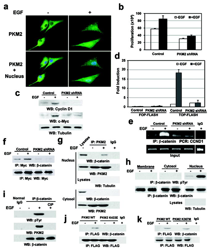
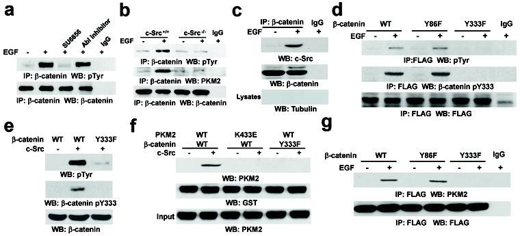
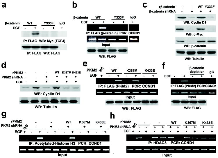
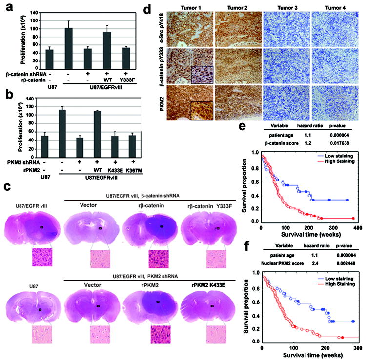
The embryonic pyruvate kinase M2 (PKM2) isoform is highly expressed in human cancer. In contrast to the established role of PKM2 in aerobic glycolysis or the Warburg effect, its non-metabolic functions remain elusive. Here we demonstrate, in human cancer cells, that epidermal growth factor receptor (EGFR) activation induces translocation of PKM2, but not PKM1, into the nucleus, where K433 of PKM2 binds to c-Src-phosphorylated Y333 of β-catenin. This interaction is required for both proteins to be recruited to the CCND1 promoter, leading to HDAC3 removal from the promoter, histone H3 acetylation and cyclin D1 expression. PKM2-dependent β-catenin transactivation is instrumental in EGFR-promoted tumour cell proliferation and brain tumour development. In addition, positive correlations have been identified between c-Src activity, β-catenin Y333 phosphorylation and PKM2 nuclear accumulation in human glioblastoma specimens. Furthermore, levels of β-catenin phosphorylation and nuclear PKM2 have been correlated with grades of glioma malignancy and prognosis. These findings reveal that EGF induces β-catenin transactivation via a mechanism distinct from that induced by Wnt/Wingless and highlight the essential non-metabolic functions of PKM2 in EGFR-promoted β-catenin transactivation, cell proliferation and tumorigenesis.


Figures

Figure 1
Figure 1. EGF induces the PKM2–β-catenin interaction in the nucleus
a, U87/EGFR cells were treated with or without EGF for 10 h. b, U87/EGFR cells with or without PKM2 depletion were plated and counted 7 days after seeding. Data represent the means ± SD of three independent experiments. c, e, U87/EGFR cells with or without PKM2 depletion were treated with or without EGF for 24 h (c) or 10 h (e). d, U87/EGFR cells with or without PKM2 depletion were transfected with TOP-FLASH or FOP-FLASH, which was followed by EGF treatment for 10 h. Data represent the means ± SD of three independent experiments. f, Myc-TCF4 was immunoprecipitated from PKM2-depleted or PKM2-undepleted U87/EGFR cells treated with or without EGF for 10 h. g, h, PKM2 (g) or β-catenin (h) was immunoprecipitated from the indicated cell fractions of U87/EGFR cells treated with or without EGF for 6 h. i, β-catenin immunoprecipitated from U87/EGFR cells with or without EGF treatment for 6 h was incubated with or without CIP (10 unit) for 30 min at 37°C followed by PBS washing for three times. j, k, U87/EGFR cells stably expressing FLAG-tagged WT PKM2, PKM2 K433E (j), or PKM2 K367M (k) were treated with or without EGF for 6 h.
Figure 2
Figure 2. c-Src phosphorylates β-catenin at Y333 upon EGFR activation
a, U87/EGFR cells were treated with SU6656 (4 μM) or an Abl inhibitor (0.2 μM) for 30 min before EGF treatment for 6 h. b, The indicated cells were treated with or without EGF for 6 h. c, β-catenin was immunoprecipitated from the nuclear fractions of U87/EGFR cells treated with or without EGF for 6 h. d, g, U87/EGFR cells transiently expressing the indicated FLAG-tagged β-catenin proteins were treated with or without EGF for 6 h. e, In vitro kinase assays were performed with purified active c-Src and purified β-catenin proteins. f, Immobilized GST-β-catenin proteins were mixed with purified His-PKM2 proteins in the presence or absence of active c-Src.
Figure 3
Figure 3. The PKM2–β-catenin interaction is required for β-catenin-induced cyclin D1 expression
a, b, U87/EGFR cells transiently expressing FLAG-β-catenin proteins were treated with or without EGF for 10 h. c, β-catenin was depleted in U87/EGFRvIII cells, followed by reconstituted expression of rβ-catenin. d, g, h, U87/EGFR cells with or without depleted PKM2 and reconstituted expression of rPKM2 were treated with or without EGF for 24 h (d) or 10 h (g, h). e, U87/EGFR cells transiently expressing the indicated FLAG-tagged PKM2 protein were treated with or without EGF for 10 h. f, U87/EGFR cells transiently expressing FLAG-PKM2 were treated with or without EGF for 10 h. β-catenin was immunoprecipitated from the cell lysates, and the remaining supernatant was used for ChIP analyses.
Figure 4
Figure 4. The PKM2–β-catenin interaction is required for tumor development
a, b, U87, U87/EGFRvIII cells with or without depleted β-catenin and reconstituted expression of rβ-catenin (a), or U87/EGFRvIII cells with or without depleted PKM2 and reconstitution of the expression of rPKM2 (b), were plated and counted 7 days after seeding. Data represent the means ± SD of three independent experiments. c, U87 (bottom left panel), U87/EGFRvIII cells with or without depleted β-catenin and reconstituted expression of rβ-catenin (top panel), or U87/EGFRvIII cells with or without depleted PKM2 and reconstituted expression of rPKM2 (bottom right panel) were intracranially injected into athymic nude mice. After two weeks, tumor growth was examined. H&E-stained coronal brain sections show representative tumor xenografts. d, IHC staining with the indicated antibodies was performed on 55 GBM specimens. Representative photos of four tumors are shown. e, f, The survival time for 84 patients with low (0-5 staining scores, blue curve) versus high (6-8 staining scores, red curve) β-catenin Y333 phosphorylation (e; low, 28 patients; high, 56 patients) and nuclear PKM2 expression (f; low, 28 patients; high, 56 patients) were compared (bottom panel). The table (top panel) shows the multivariate analysis after adjustment for patient age, indicating the significance level of the association of Y133-phosphorylated β-catenin expression (e) or nuclear PKM2 expression (f) with patient survival. Empty circles represent the deceased patients, and filled circles represent the censored (alive at last clinical follow-up) patients.

Comment in

References

    1. Vander Heiden MG, Cantley LC, Thompson CB. Understanding the Warburg effect: the metabolic requirements of cell proliferation. Science. 2009;324:1029–1033. - PMC - PubMed
    1. Cairns RA, Harris IS, Mak TW. Regulation of cancer cell metabolism. Nat Rev Cancer. 2011;11:85–95. - PubMed
    1. Koppenol WH, Bounds PL, Dang CV. Otto Warburg’s contributions to current concepts of cancer metabolism. Nat Rev Cancer. 2011;11:325–337. - PubMed
    1. Lu Z, Hunter T. Wnt-independent beta-catenin transactivation in tumor development. Cell Cycle. 2004;3:571–573. - PubMed
    1. Lu Z, Jiang G, Blume-Jensen P, Hunter T. Epidermal growth factor-induced tumor cell invasion and metastasis initiated by dephosphorylation and downregulation of focal adhesion kinase. Mol Cell Biol. 2001;21:4016–4031. - PMC - PubMed

Publication types