. 2011 Nov 6;480(7378):547-51.
doi: 10.1038/nature10648.

Dopamine neurons derived from human ES cells efficiently engraft in animal models of Parkinson's disease

Affiliations

Dopamine neurons derived from human ES cells efficiently engraft in animal models of Parkinson's disease

Sonja Kriks et al. Nature. .

Abstract

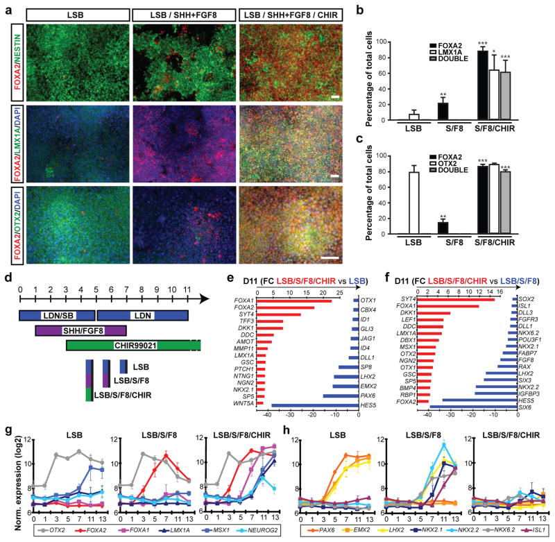
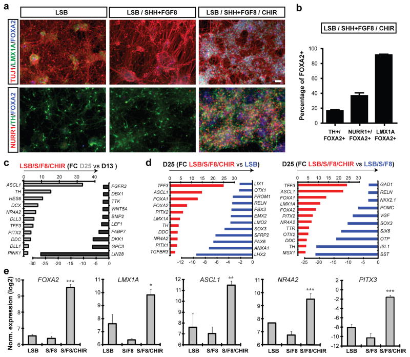
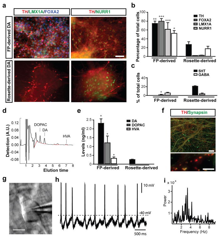
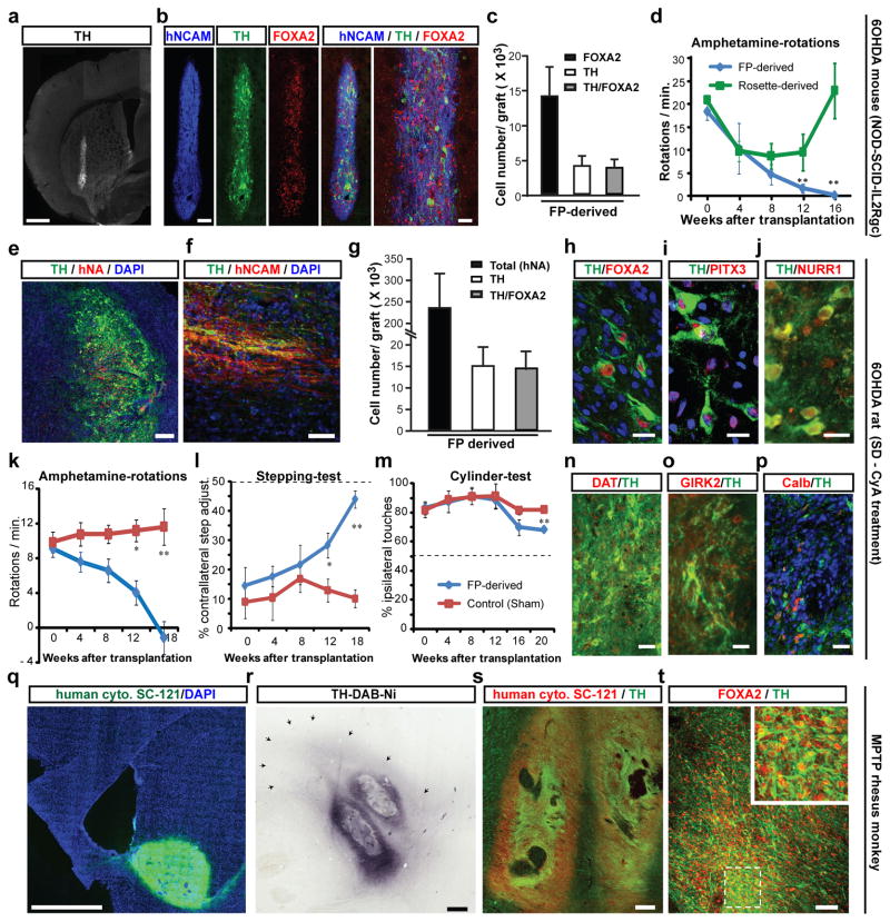
Human pluripotent stem cells (PSCs) are a promising source of cells for applications in regenerative medicine. Directed differentiation of PSCs into specialized cells such as spinal motoneurons or midbrain dopamine (DA) neurons has been achieved. However, the effective use of PSCs for cell therapy has lagged behind. Whereas mouse PSC-derived DA neurons have shown efficacy in models of Parkinson's disease, DA neurons from human PSCs generally show poor in vivo performance. There are also considerable safety concerns for PSCs related to their potential for teratoma formation or neural overgrowth. Here we present a novel floor-plate-based strategy for the derivation of human DA neurons that efficiently engraft in vivo, suggesting that past failures were due to incomplete specification rather than a specific vulnerability of the cells. Midbrain floor-plate precursors are derived from PSCs 11 days after exposure to small molecule activators of sonic hedgehog (SHH) and canonical WNT signalling. Engraftable midbrain DA neurons are obtained by day 25 and can be maintained in vitro for several months. Extensive molecular profiling, biochemical and electrophysiological data define developmental progression and confirm identity of PSC-derived midbrain DA neurons. In vivo survival and function is demonstrated in Parkinson's disease models using three host species. Long-term engraftment in 6-hydroxy-dopamine-lesioned mice and rats demonstrates robust survival of midbrain DA neurons derived from human embryonic stem (ES) cells, complete restoration of amphetamine-induced rotation behaviour and improvements in tests of forelimb use and akinesia. Finally, scalability is demonstrated by transplantation into parkinsonian monkeys. Excellent DA neuron survival, function and lack of neural overgrowth in the three animal models indicate promise for the development of cell-based therapies in Parkinson's disease.


Figures

Figure 1
Figure 1. Induction and neurogenic conversion of hESC-derived midbrain FP precursors is dependent on CHIR99021 addition
a) Immunocytochemistry at day 11 for FOXA2 (red), NESTIN (green, upper panels), LMX1A (green, middle panels) and OTX2 (green, lower panels). b,c) Quantification of the data presented in (a); mean ± SEM, n=3 (independent experiments): *** p < 0.001; ** p < 0.01; p < 0.05 (compared to LSB, Dunnett test). d) Diagram of culture conditions. e,f) Selected lists of differentially expressed transcripts at day 11 comparing LSB/S/F8/CHIR versus LSB (e) or versus LSB/S/F8 (f). g,h) Temporal gene expression analysis for markers of midbrain DA precursor (g), forebrain and ventral non-DA precursor identity (h). Scale bars: 50 μm.
Figure 2
Figure 2
Immunocytochemical and molecular analysis of midbrain DA neuron fate in LSB/S/F8/CHIR treated versus LSB/S/F8 (hypothalamic) and forebrain LSB (dorsal forebrain) fates. a) Immunocytochemistry at day 25 for co-expression of FOXA2 (blue) with Tuj1(red)/LMX1A(green) (upper panels) and NURR1(red)/TH(green) (lower panels). b) Quantitative co-expression analysis for LSB/S/F8/CHIR; mean ± SEM, n=3 (independent experiments). c,d) Global gene expression analysis at day 25 (triplicates each). Selected lists of differentially expressed transcripts comparing day 13 versus day 25 in LSB/S/F8/CHIR (c) LSB/S/F8/CHIR versus LSB (d, left panel) and LSB/S/F8 (d, right panel). e) Gene expression analysis for key midbrain DA neuron markers. Significance compared to LSB: Dunnett test: *** p < 0.001; ** p < 0.01; * p < 0.05). Scale bars: 50 μm.
Figure 3
Figure 3. In vitro maturation and functional characterization of FP versus rosette-derived midbrain DA neurons
a) Immunocytochemistry at day 50 for TH (red), with LMX1A (green) and FOXA2 (blue; left panels) and NURR1 (green, right panels). b) Quantification of TH+, FOXA2+, LMX1+ and NURR1+ cells in rosette- versus FP-derived (LSB/S/F8/CHIR) cultures. c) Quantification of serotonin+ (5-HT), and GABA+ neuronal subtypes at day 50 in rosette-versus FP-derived cultures. d,e) HPLC analysis for DA and metabolites d) Representative HPLC chromatogram in a sample of FP-derived cultures. e) Levels of DA, DOPAC and HVA in FP-and rosette-derived cultures. f) Immunocytochemistry in FP-derived cultures (day 80) for TH (red) and synapsin (green). g-i) Electrophysiological analyses of FP cultures at day 80. Phase contrast image of a patched neuron (g) and corresponding recordings (h). i) Power analysis showing membrane potential oscillations characteristic of DA neuron identity (2~5Hz) Mean ± SEM; significance (panels b, c, e) comparing FP versus rosette-derived cultures: Student’s T-test: *** p < 0.001; ** p < 0.01; p < 0.05). Scale bars: 50 μm in (a), 20 μm in (f, upper panel), 5 μm in (f, lower panel) and 20 μm in (g)
Figure 4
Figure 4. In vivo survival and function of FP-derived human DA neurons in mouse, rat and monkey PD hosts
a-d) 6-OHDA lesioned adult mice (NOD-SCID IL2Rgc null strain): a) TH expression and graft morphology at 4.5 months after transplantation. b) Expression of human specific marker (hNCAM, blue), TH (green), and FOXA2 (red). c) Quantification of FOXA2+ and TH+ cells in FP-derived grafts (mean ± SEM, n=4 at 4.5 months post grafting). d) Amphetamine-induced rotation analysis in FP- (blue) versus rosette-derived (green) grafts. Scale bars: 500 μm in (a), and 100 and 40 μm in (b). e-p) 6-OHDA lesioned adult rats: Immunohistochemistry for TH (green) and human specific markers (red) hNA (e) and hNCAM (f). g) Stereological quantification of hNA+, TH+ and TH+ cells co-expressing FOXA2 (average graft volume = 2.6 ± 0.6 mm3). h-j) Co-expression of TH (green) with FOXA2, PITX3 and NURR1 (red). k-m) Behavioral analysis in FP- versus sham-grafted animals. k) Amphetamine-induced rotational asymmetry. l) stepping test: measuring forelimb akinesia in affected versus non-affected side. m) Cylinder test: measuring ipsi- versus contra-lateral paw preference. Grafted animals showed significant improvement in all three tests (p < 0.01 at 4.5–5 month; n=4–6 each). n-p) Immunohistochemistry for TH (green) and co-expression (red) with DAT (n), GIRK2 (o) and calbindin (p). Significance levels (panels d, k, l, m): ** p < 0.01; p < 0.05). Scale bars: 200 μm in (e), 50 μm in (f), 20 μm in (h-j) and 40 μm in (n-p). q-t) Adult MPTP lesioned rhesus monkeys. q) Representative graft at 1 month after transplantation expressing human specific cytoplasm marker SC-121 (green). r) TH expression in graft with surrounding TH+ fibers (arrows). s) Co-expression of SC-121 (red) and TH (green). t) Co-expression of FOXA2 (red) and TH+ (green). Scale bars: 2mm for (q), 500 μm for (r), 200 μm for (s), and 50 μm for (t).

Comment in

Similar articles

Cited by

References

    1. Li XJ, et al. Specification of motoneurons from human embryonic stem cells. Nat Biotechnol. 2005;23:215–221. - PubMed
    1. Perrier AL, et al. From the Cover: Derivation of midbrain dopamine neurons from human embryonic stem cells. Proc Natl Acad Sci U S A. 2004;101:12543–8. - PMC - PubMed
    1. Tabar V, et al. Therapeutic cloning in individual Parkinsonian mice. Nature Med. 2008;14:379–381. - PMC - PubMed
    1. Wernig M, et al. Neurons derived from reprogrammed fibroblasts functionally integrate into the fetal brain and improve symptoms of rats with Parkinson’s disease. Proc Natl Acad Sci U S A. 2008;105:5856–5861. - PMC - PubMed
    1. Lindvall O, Kokaia Z. Stem cells in human neurodegenerative disorders--time for clinical translation? J Clin Invest. 2010;120:29–40. - PMC - PubMed

Publication types

Associated data