Review
. 2011 Dec;15(6):845-52.
doi: 10.1016/j.cbpa.2011.10.008. Epub 2011 Nov 5.

Single-molecule analysis of telomerase structure and function

Affiliations
Review

Single-molecule analysis of telomerase structure and function

Martin Hengesbach et al. Curr Opin Chem Biol. 2011 Dec.

Abstract

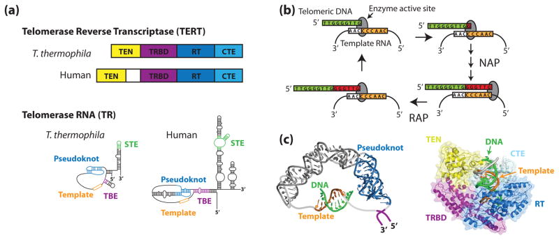
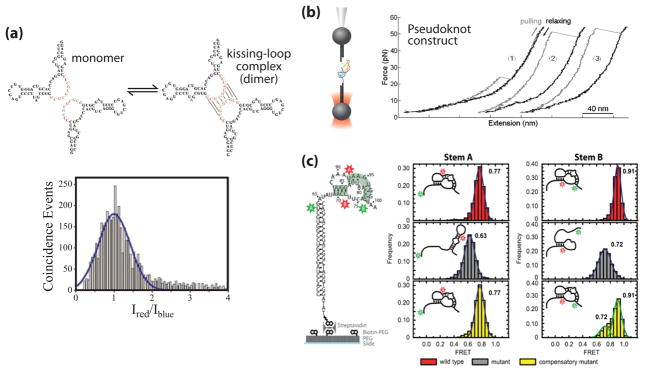
The telomerase ribonucleoprotein is a specialized reverse transcriptase required to maintain protective chromosome end-capping structures called telomeres. In most cells, telomerase is not active and the natural shortening of telomeres with each round of DNA replication ultimately triggers cell growth arrest. In contrast, the presence of telomerase confers a high level of renewal capacity upon rapidly dividing cells. Telomerase is aberrantly activated in 90% of human cancers and thus represents an important target for anticancer therapeutics. However, the naturally low abundance of telomerase has hampered efforts to obtain high-resolution models for telomerase structure and function. To circumvent these challenges, single-molecule techniques have recently been employed to investigate telomerase assembly, structure, and catalysis.


Figures

Figure 1
Figure 1. The telomerase ribonucleoprotein complex
(a) Tetrahymena thermophila and human TERTs and TRs share a similar domain organization. In both species, TERT is composed of an N-terminal (TEN) domain, an RNA-binding domain (TRBD), a reverse transcriptase domain (RT), and a C-terminal extension (CTE). Conserved RNA structural features include an RNA pseudoknot fold, template, template boundary element (TBE), and a stem terminal element (STE). (b) During the telomerase catalytic cycle, telomeric DNA is positioned in the enzyme active site by basepairing alignment with the template RNA. Individual nucleotides are reverse transcribed off of the template RNA in a process known as nucleotide addition processivity (NAP). When the end of the template is reached, the DNA-RNA duplex is denatured and the RNA is re-positioned to add another repeat, a process known as repeat addition processivity (RAP). (c) In silico modeling of human TR (left) and human TERT (right) (adapted from [1,2] with permission). Zhang and co-workers used residual dipolar coupling orientation constraints to model the core region of human TR using existing NMR structures. Steczkiewicz et al. used homology modeling and structure prediction algorithms to model human TERT based on solved structures of the Tribolium castaneum TERT and the Tetrahymena thermophila TEN and TRBD domains, and used molecular docking to predict the position of the TEN domain within the enzyme.
Figure 2
Figure 2. Single molecule approaches to telomerase research
(a) Two-color coincidence detection (TCCD). In the TCCD approach, two partially overlapping red and blue lasers are focused through a confocal microscope and very dilute biological macromolecules labeled with either a blue or red dye are studied. Transient diffusion of dye-labeled molecules through the confocal volume gives rise to fluorescence bursts (lower panel), and the ratio of red and blue fluorescence intensities provides information about the composition and stoichiometry of individual complexes. (b) Single molecule FRET measured by prism-type total internal reflection fluorescence (TIRF) microscopy. A molecule of interest is labeled with a FRET donor (green) and acceptor (red) dye and surface immobilized via a biotin-streptavidin linkage onto a microscope slide. The sample is illuminated by the evanescent wave generated by TIRF microscopy, which suppresses fluorescence background to levels that permit prolonged detection of individual FRET pairs. Typically, FRET is measured as the ratio of the fluorescence intensity of the acceptor dye divided by the sum of the donor plus acceptor intensities. (c) Force-measuring optical trap. The RNA molecule of interest is attached to a micron-scale bead held on a glass micropipette as well as a second bead held in the optical trap. Displacement of the micropipette away from the optical trap results in a stretching force applied to the RNA molecule, and both the applied force and molecular displacements are measured in real time.
Figure 3
Figure 3. Single molecule studies of telomerase RNA structure
(a) Proposed dimerization interaction in the J7b/8a region of the CR7 region of hTR (adapted from [3] with permission). RNA is 5′ end labeled with either Alexa 488 or Alexa 647 in a 1:1 ratio and diluted to a total concentration of 100 pM. A histogram of the log ratio of red to blue fluorescence for single molecules diffusing through the excitation volume was indicative of a single distribution centered at a 1:1 ratio, corresponding to the hTR-Alexa 488: hTR-Alexa 647 dimer. (b) Mechanical folding/unfolding of the hTR pseudoknot domain RNA (adapted from [4] with permission). The pseudoknot construct used in this study was oriented between two DNA handles and held in an optical trap. The RNA molecule is pulled (grey line) and relaxed (black line) at a constant rate of 100 nm/sec as the resultant force is measured. The rip at ~24 pN (trace 1) corresponds to the unfolding force of an alternative hairpin conformation observed in a subset of traces. The rips at ~50 pN (traces 2 & 3) correspond to the unfolding force of the complete pseudoknot, resulting in a rip size of ~36 nm which corresponds to the dimensions of the pseudoknot. (c) Single molecule FRET (smFRET) studies of the Tetrahymena TR (tTR) pseudoknot domain RNA (adapted from [5] with permission). Constructs were labeled with a donor dye (green) at residue U63 and an acceptor dye (red) at U92 to measure folding of stem A or labeled with a donor dye at U73 and an acceptor dye at U99 to measure folding of stem B. smFRET histograms were obtained with the stem A and stem B labeling sites for the wild-type RNA sequence (red), a mutant pseudoknot designed to disrupt basepairing in stem A (grey), and a compensatory mutant designed to restore basepairing (yellow).
Figure 4
Figure 4. Single molecule analysis of telomerase RNP assembly, composition, and activity
(a) Schematic diagram of proposed p65-mediated Tetrahymena telomerase RNP assembly pathway. For smFRET measurements, donor (green) and acceptor (red) dyes were placed on tTR to monitor RNA folding during the RNP assembly process. A representative smFRET trace displays a FRET value of 0.29 in the absence of proteins. Upon addition of p65 and TERT (dashed line), the RNA undergoes stepwise folding transitions (black arrows) to 0.46 FRET and 0.65 FRET, corresponding to the sequential binding of p65 and TERT, respectively (adapted from [6] with permission). (b) Characterization of telomerase composition by TCCD. TCCD histograms for human telomerase RNP complexes harboring blue and red dyes on either: hTERT and hTR (left); hTERT and DNA substrate (middle); or hTR and DNA substrate (right) are consistent with a 1:1:1 stoichiometry of hTERT:hTR:DNA substrate within the active telomerase complex (adapted from [7] with permission). (c) Human telomerase activity detection by TCCD. Reconstituted human telomerase is incubated with telomeric DNA primers labeled with a blue reference dye and reacted in the presence dATP coupled to a red dye. One dATP is incorporated for each telomeric repeat, thus the ratio of blue and red fluorescence intensities provides a direct measure of the number of repeats added to each DNA substrate (adapted from [8] with permission). (d) smFRET-based Tetrahymena telomerase structure-function assay. Telomeric DNA primers were surface immobilized and incubated with RNP complexes reconstituted with tTR harboring FRET donor and acceptor dyes. Individual smFRET measurements were made during transient telomerase binding events. During the activity detection phase, DNA primers were labeled in situ with a new FRET donor (green star) and telomerase activity was detected by measuring the hybridization kinetics of an acceptor (red star) labeled detection oligo (DO) (adapted from [9] with permission).

References

    1. Palm W, de Lange T. How shelterin protects mammalian telomeres. Annu Rev Genet. 2008;42:301–334. - PubMed
    1. Greider CW, Blackburn EH. A telomeric sequence in the RNA of Tetrahymena telomerase required for telomere repeat synthesis. Nature. 1989;337:331–337. - PubMed
    1. Lingner J, Hughes TR, Shevchenko A, Mann M, Lundblad V, Cech TR. Reverse transcriptase motifs in the catalytic subunit of telomerase. Science. 1997;276:561–567. - PubMed
    1. Collins K. The biogenesis and regulation of telomerase holoenzymes. Nat Rev Mol Cell Biol. 2006;7:484–494. - PMC - PubMed
    1. Gillis AJ, Schuller AP, Skordalakes E. Structure of the Tribolium castaneum telomerase catalytic subunit TERT. Nature. 2008;455:633–637. - PubMed

Publication types

MeSH terms

LinkOut - more resources