Automatic analysis of EMG during clonus
- PMID: 22057220
- PMCID: PMC3249492
- DOI: 10.1016/j.jneumeth.2011.10.017
Automatic analysis of EMG during clonus
Abstract
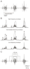
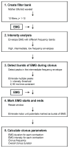
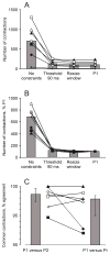
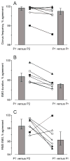
Clonus can disrupt daily activities after spinal cord injury. Here an algorithm was developed to automatically detect contractions during clonus in 24h electromyographic (EMG) records. Filters were created by non-linearly scaling a Mother (Morlet) wavelet to envelope the EMG using different frequency bands. The envelope for the intermediate band followed the EMG best (74.8-193.9 Hz). Threshold and time constraints were used to reduce the envelope peaks to one per contraction. Energy in the EMG was measured 50 ms either side of each envelope (contraction) peak. Energy values at 5% and 95% maximal defined EMG start and end time, respectively. The algorithm was as good as a person at identifying contractions during clonus (p=0.946, n=31 spasms, 7 subjects with cervical spinal cord injury), and marking start and end times to determine clonus frequency (intra class correlation coefficient, α: 0.949), contraction intensity using root mean square EMG (α: 0.997) and EMG duration (α: 0.852). On average the algorithm was 574 times faster than manual analysis performed independently by two people (p ≤ 0.001). This algorithm is an important tool for characterization of clonus in long-term EMG records.
Copyright © 2011 Elsevier B.V. All rights reserved.
Figures


References
-
- Adams MM, Hicks AL. Spasticity after spinal cord injury. Spinal Cord. 2005;43:577–86. - PubMed
-
- Beres-Jones JA, Johnson TD, Harkema SJ. Clonus after human spinal cord injury cannot be attributed solely to recurrent muscle-tendon stretch. Exp Brain Res. 2003;149:222–36. - PubMed
-
- Bonato P, D’Alessio T, Knaflitz M. A statistical method for the measurement of muscle activation intervals from surface myoelectric signal during gait. IEEE Trans Biomed Eng. 1998;45:287–99. - PubMed
-
- Cook WA., Jr Antagonistic muscles in the production of clonus in man. Neurology. 1967;17:779–96. - PubMed
-
- Di Fabio RP. Reliability of computerized surface electromyography for determining the onset of muscle activity. Phys Ther. 1987;67:43–8. - PubMed
Publication types
MeSH terms
Substances
Grants and funding
LinkOut - more resources
Full Text Sources
Medical

