. 2011 Nov 6;12(12):1176-83.
doi: 10.1038/ni.2157.

The E3 ligase Itch and deubiquitinase Cyld act together to regulate Tak1 and inflammation

Affiliations

The E3 ligase Itch and deubiquitinase Cyld act together to regulate Tak1 and inflammation

Neesar Ahmed et al. Nat Immunol. .

Abstract

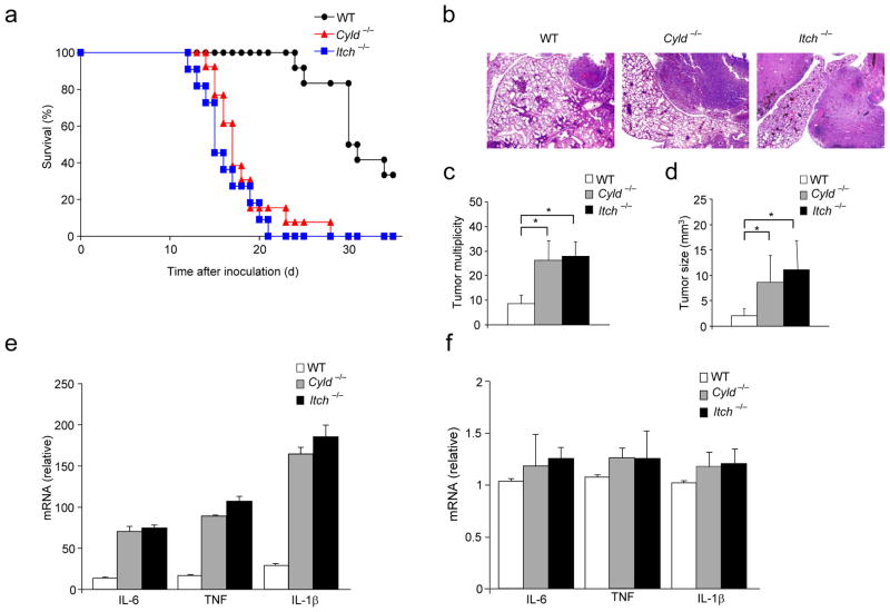
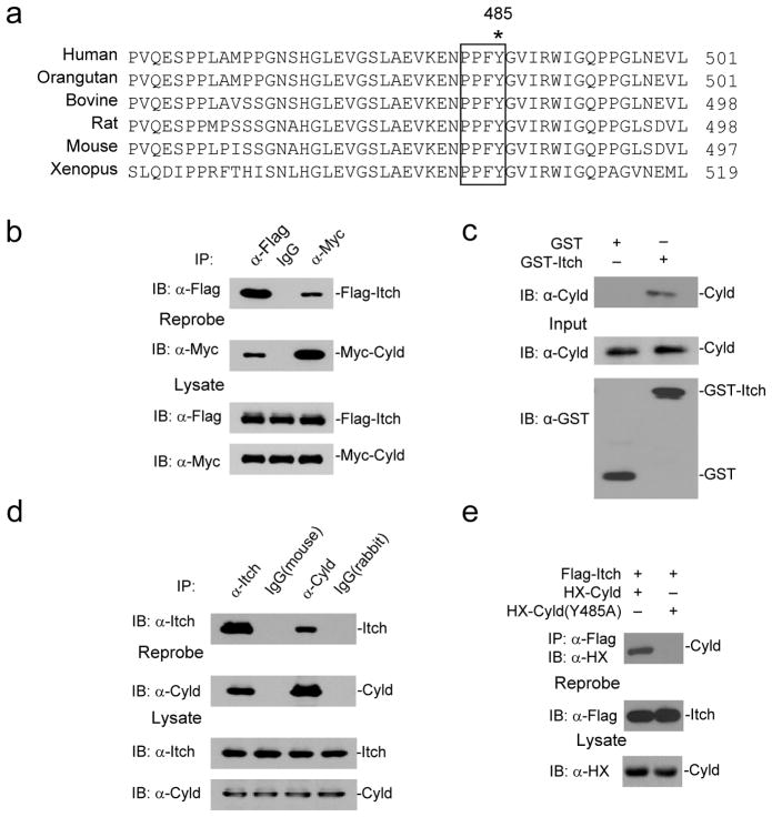
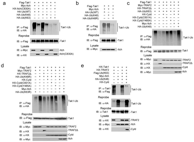
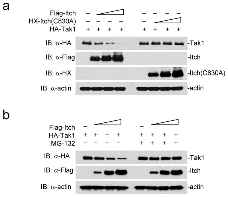
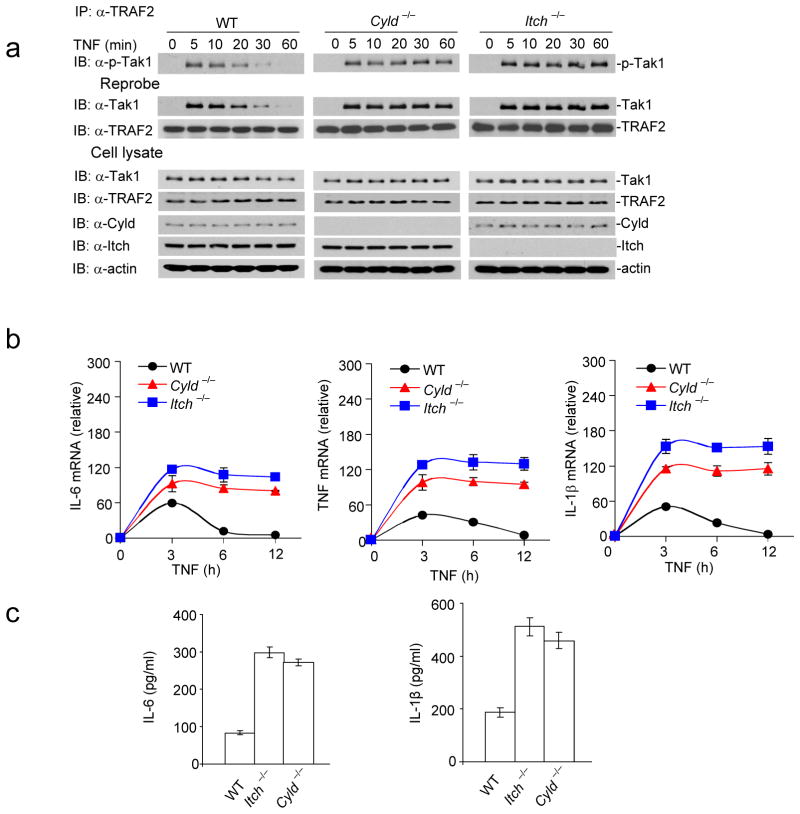
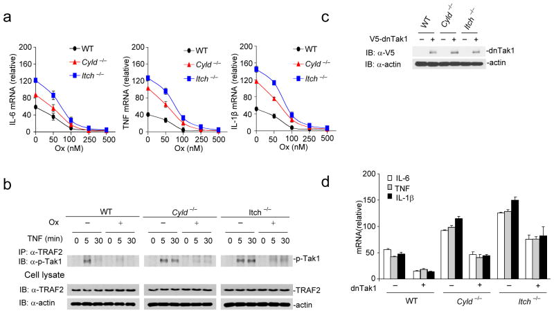
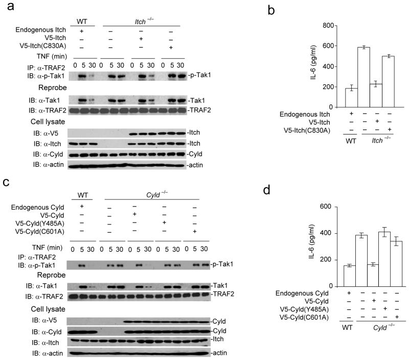
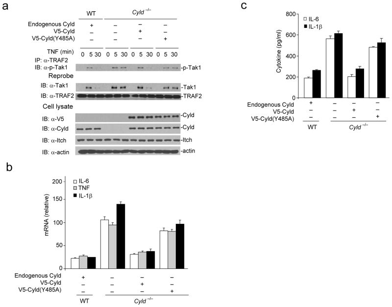
Chronic inflammation has been strongly associated with tumor progression, but the underlying mechanisms remain elusive. Here we demonstrate that E3 ligase Itch and deubiquitinase Cyld formed a complex via interaction through 'WW-PPXY' motifs. The Itch-Cyld complex sequentially cleaved Lys63-linked ubiquitin chains and catalyzed Lys48-linked ubiquitination on the kinase Tak1 to terminate inflammatory signaling via tumor necrosis factor. Reconstitution of wild-type Cyld but not the mutant Cyld(Y485A), which cannot associate with Itch, blocked sustained Tak1 activation and proinflammatory cytokine production by Cyld(-/-) bone marrow-derived macrophages. Deficiency in Itch or Cyld led to chronic production of tumor-promoting cytokines by tumor-associated macrophages and aggressive growth of lung carcinoma. Thus, we have identified an Itch-Cyld-mediated regulatory mechanism in innate inflammatory cells.


Conflict of interest statement

Competing financial interests

The authors declare no competing financial interests.

Figures

Figure 1
Figure 1
Itch−/− and Cyld−/− mice develop elevated growth and metastasis of Lewis Lung Carcinoma. (a) % survival of WT (n=12), Cyld−/− (n=13) and Itch−/−(n=11) mice inoculated with 1×10⁶ Lewis Lung Carcinoma (LLC) cells via tail vein. (b) Representative H&E stained histological sections of the lungs of wild type, Itch−/− and Cyld−/− mice on day 15 after inoculation of LLC cells (1×10⁶ cells). (c) Lung tumor multiplicity and (d) Tumor size. (e) Cytokine mRNA levels in CD11b+ macrophages from lung tumors of WT, Itch−/− and Cyld−/− mice inoculated with LLC cells (1×10⁶) via tail vein. (f) Cytokine abundance in naïve Itch−/− and Cyld−/− lung tissues. *P<0.01(paired t-test). Data are representative of three independent experiments.
Figure 2
Figure 2
Itch and Cyld form a complex via interaction through PPXY motif. (a) Sequence alignment of PPXY motif of Cyld which is conserved across species. Y485 critical for interaction with Itch is indicated (*). (b) 293T cells transiently co-transfected with expression vectors for Flag-Itch and Myc-Cyld. The cell lysate was immunoprecipitated using mouse IgG, antibodies against Flag and Myc. The immunoprecipitates were blotted using antibodies against Flag and Myc. (c) Immunoblot of His-Cyld precipitated using GST or GST-Itch purified from E. coli. The input controls for His-Cyld, GST and GST-Itch are shown below (d) BMDMs were stimulated with TNF for thirty minutes. The cell lysates were immunoprecipitated using mouse IgG, rabbit IgG, antibodies against Itch and Cyld. The immunoprecipitates were analyzed by immunoblot with antibodies against Itch and Cyld. (e) 293T cells were co-transfected with expression vectors for HX-Cyld(Y485A) and Flag-Itch. The cells lysate was immunoprecipitated using antibody against Flag and immunoblotted using antibody against HX. Data are representative of three (b, c and e) and two (d) experiments.
Figure 3
Figure 3
Itch and Cyld sequentially cleave K63-linked ubiquitination and catalyze K48-linked ubiquitination to deactivate Tak1. (a) 293T cells co-transfected with Flag-Tak1 along with Myc-Itch, HX-Itch (C830A), HA tagged Ub(WT), Ub(K63) and Ub(K48) as indicated. The cell lysate was immunoprecipitated using antibody against Flag and immunoblotted using antibody against HA. (b) 293T cells co-transfected with Flag-Tak1 along with Myc-Itch, HA-Ub(WT), HA-Ub(K48) and Ub(K48R) as indicated. The cell lysate was immunoprecipitated using antibody against Flag and immunoblotted using antibody against HA. (c) 293T cells co-transfected with Flag-Tak1 along with Myc-TRAF2, HX-TRAF2Δ, HA-Ub(K63), HX-Cyld, HX-Cyld(C601A), HX-Cyld(Y485A), Myc-Itch, HA-Ub(K48) in various combinations as indicated. The cell lysate was immunoprecipitated using antibody against Flag and immunoblotted using antibody against HA. (d) 293T cells were transfected as in (b) utilizing Ub(K48R) and Ub(K63R). (e) HX-Tak1, HX-TRAF2 and Flag-Ub(K63), HA-Ub(K48), Myc-Itch, and HX-Cyld were co-expressed in 293T cells. The cell lysate was immunoprecipitated using antibody against Tak1 and immunoblotted with antibody against Flag to detect K63-linked ubiquitination. The membranes were reprobed with antibody against HA to detect K48-linked ubiquitination. Data are representative of three or more independent experiments.
Figure 4
Figure 4
Itch-mediated polyubiquitination results in Tak1 degradation. (a) 293T cells were co-transfected with HA-Tak1 (1μg) and increasing concentrations of Flag-Itch or Itch(C830A). The cellular Tak1 level was analyzed by immunoblotting the lysate using antibody against HA. The level of expression of Itch and Itch-C830A was analyzed by reprobing the membranes using antibodies against Flag and HX. (b) 293T cells were transfected with HA-Tak1 (1μg) and increasing concentrations of Flag-Itch, the cells were cultured in the presence or absence of MG132. The cellular Tak1 level was analyzed by immunoblotting the lysate using antibody against HA. Data are representative of two experiments.
Figure 5
Figure 5
Sustained Tak1 activation results in chronic production of inflammatory cytokines by Itch−/− and Cyld−/− BMDMs. (a) Itch−/− and Cyld−/− BMDMs were stimulated with TNF and lysed at the indicated time points. The lysate was immunoprecipitated with antibody against TRAF2 and immunoblotted with antibody against p-Tak1. The membranes were reprobed with antibodies against Tak1 and TRAF2. The total cell lysate was blotted with antibodies against Tak1, TRAF2, Cyld, Itch and actin. (b) BMDMs were stimulated with TNF, total RNA was isolated at the indicated time points and expression of IL-6, TNF and IL-1β was analyzed using real-time PCR. (c) IL-6 and IL-1β concentration were assayed in the culture supernatant by ELISA. Data are representative of three experiments (b–c mean and s.d. of triplicate wells).
Figure 6
Figure 6
Inhibiting Tak1 activation rescues elevated inflammatory cytokine production by Itch−/− and Cyld−/− BMDMs (a) BMDMs were pre-incubated with indicated amounts of (5Z)-7-Oxozeaenol(Ox) and stimulated with TNF for 6 hours, expression of IL-6, TNF, and IL-1β was analyzed by real-time PCR. (b) BMDMs were pretreated with 100 nM of Ox for 30 minutes and then stimulated with TNF for the indicated time points. The cell lysates were immunoprecipitated using antibody against TRAF2 and immunoblotted using antibody against p-Tak1. The total lysate was immunoblotted using antibodies against TRAF2 and actin. (c) WT, Itch−/− and Cyld−/− BMDMs were transduced with a lentiviral vector encoding dominant negative Tak1 (dnTak1). The efficiency of ectopic expression was analyzed by immunoblotting the total cell lysate using antibody against V5. (d) WT, Itch−/− and Cyld−/− BMDMs transduced with dnTak1 were stimulated with TNF and expression of IL-6, TNF and IL-1β was analyzed by real-time PCR. Data are representative of three experiments. (a, d mean and s.d. of triplicate wells).
Figure 7
Figure 7
Ligase activity of Itch, DUB activity of Cyld, and Cyld-Itch interaction is necessary for termination of TNF induced Tak1 activation. (a) Itch−/− MEFs were reconstituted with either V5-Itch(WT) or V5-Itch(C830A) mutant using a lentiviral transduction system. The cells lysed at indicated time points post TNF stimulation. The lysate was immunoprecipitated using against TRAF2 and immunoblotted with antibody against p-Tak1. The same membrane was reprobed with antibody against Tak1. The cell lysate from WT MEFs stimulated with TNF was used as control. (b) Itch−/− MEFs transduced with V5-Itch(WT) or V5-Itch(C830A) mutant as in (a) were stimulated with TNF and expression of IL-6 was analyzed by ELISA in the culture supernatant. (c) Cyld−/− MEFs were reconstituted with V5-Cyld(WT), V5-Cyld(C601A) or V5-Cyld(Y485A) mutant. The cells lysed at indicated time points post TNF stimulation. The lysate was immunoprecipitated using antibody against TRAF2 and immunoblotted with antibody against p-Tak1. The same membrane was reprobed with antibody against Tak1. (d) Cyld−/− MEFs transduced with V5-Cyld(WT), V5-Cyld(Y485A) or V5-Cyld(C601A) mutant as in (c) stimulated with TNF and expression of IL-6 was analyzed by ELISA in the culture supernatant. Data are representative of three (b, d) and two (a, c) experiments. (b,d mean and s.d. of triplicate wells).
Figure 8
Figure 8
Reconstitution of Cyld−/− BMDMs with WT but not Cyld(Y485A) mutant rescues defects in termination of Tak1 activation and chronic production of inflammatory cytokines. (a) V5-Cyld(WT) or V5-Cyld(Y485A) mutant was ectopically expressed in Cyld−/− BMDMs using a lentiviral transduction system. The cells lysed at indicated time points post TNF stimulation. The lysate was immunoprecipitated using antibody against TRAF2 and immunoblotted with antibody against p-Tak1. The same membrane was reprobed with antibody against Tak1. The cell lysate from WT BMDMs stimulated with TNF was used as control in these experiments. (b) WT and Cyld−/− transduced with V5- Cyld(WT) or V5-Cyld(Y485A) mutant were stimulated with TNF and expression of IL-6, TNF and IL-1β was analyzed by real-time PCR. (c) IL-6 and IL-1β concentration were assayed in the culture supernatant by ELISA. Data are representative of at least three independent experiments (b–c mean and s.d. of triplicate wells).

Comment in

References

    1. Grivennikov SI, Greten FR, Karin M. Immunity, inflammation, and cancer. Cell. 2010;140:883–899. - PMC - PubMed
    1. Mantovani A, Allavena P, Sica A, Balkwill F. Cancer-related inflammation. Nature. 2008;454:436–444. - PubMed
    1. Takahashi H, Ogata H, Nishigaki R, Broide DH, Karin M. Tobacco smoke promotes lung tumorigenesis by triggering IKKbeta- and JNK1-dependent inflammation. Cancer Cell. 17:89–97. - PMC - PubMed
    1. Moghaddam SJ, et al. Promotion of lung carcinogenesis by chronic obstructive pulmonary disease-like airway inflammation in a K-ras-induced mouse model. Am J Respir Cell Mol Biol. 2009;40:443–453. - PMC - PubMed
    1. Van den Heuvel MM, Burgers SA, van Zandwijk N. Immunotherapy in non-small-cell lung carcinoma: from inflammation to vaccination. Clin Lung Cancer. 2009;10:99–105. - PubMed

Publication types

MeSH terms