. 2011 Nov 6;17(12):1680-4.
doi: 10.1038/nm.2555.

Intra-arterial catheter for simultaneous microstructural and molecular imaging in vivo

Affiliations

Intra-arterial catheter for simultaneous microstructural and molecular imaging in vivo

Hongki Yoo et al. Nat Med. .

Abstract

Advancing understanding of human coronary artery disease requires new methods that can be used in patients for studying atherosclerotic plaque microstructure in relation to the molecular mechanisms that underlie its initiation, progression and clinical complications, including myocardial infarction and sudden cardiac death. Here we report a dual-modality intra-arterial catheter for simultaneous microstructural and molecular imaging in vivo using a combination of optical frequency domain imaging (OFDI) and near-infrared fluorescence (NIRF) imaging. By providing simultaneous molecular information in the context of the surrounding tissue microstructure, this new catheter could provide new opportunities for investigating coronary atherosclerosis and stent healing and for identifying high-risk biological and structural coronary arterial plaques in vivo.


Figures

Figure 1
Figure 1
Schematic of the dual-modality intra-arterial catheter for simultaneous microstructural and molecular imaging using optical frequency-domain imaging (OFDI) and near-IR fluorescence (NIRF). OFDI and NIRF systems are combined in one system by a dual-modality rotary junction that rotates and pulls back the imaging probe contained within the transparent catheter sheath, in a manner analogous to that used in standalone OCT, OFDI and intravascular ultrasound (IVUS). The imaging probe consists of a double-clad fiber that transmits OFDI and NIRF light through separate concentric light-guiding channels and focuses the beams into the sample. In the OFDI system, the light source changes its wavelength rapidly as a function of time. The depth profile (A-line) is reconstructed by taking the Fourier transform of the spectral interference signal that is generated by combining the reference and back-reflected signals from tissue microstructures. Depth profiles are continuously acquired to form cross-sectional images while the probe is rotating. NIR fluorescence emission, which reports molecular information, is also simultaneously acquired at every A-line acquisition. Scale bars, 100 µm.
Figure 2
Figure 2
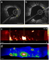
Images of a cadaveric human coronary artery with a stent containing Cy7-labeled fibrin in vitro. (a) OFDI cross-sectional image with thrombus (yellow asterisks). (b) OFDI cross-sectional image with stent struts and micro-thrombi (yellow asterisk). (c) NIRF cylindrical rendering of fibrin signal acquired by the dual-modality imaging catheter. (d) Fluorescence reflectance imaging (FRI) with Cy7 filter set. The FRI results strongly correspond to the dual modality OFDI-NIRF image. Scale bars, 1 mm.
Figure 3
Figure 3
Dual-modality OFDI-NIRF images of an iliac artery of a rabbit with an implanted NIR fluorescent fibrin-coated stent, in vivo. (a) A representative cross-sectional image of OFDI data. OFDI shows the cross-sectional arterial microscopic architectural morphology, including thrombus (red arrow) and stent struts (yellow asterisks). (b) A corresponding one-dimensional signal of NIRF data simultaneously acquired with OFDI. The NIRF signal shows strong peaks where the thrombi were detected in OFDI (red arrows in a,b). The NIRF signal was negligible in areas that did not show OFDI evidence of thrombus or fibrin by histology (blue arrowhead in a,b) (c) OFDI (gray scale) with thrombus segmentation (purple) (left column), overlaid OFDI (gray scale) and NIRF (yellow scale) images (middle column), and corresponding H&E histology (Original magnification ×25) (right column). The top row and the insets demonstrate OFDI, NIRF, and histology data showing corresponding microstructural features, such as a side branch (red asterisk), stent struts (yellow asterisks in OFDI, black asterisks in H&E), and thrombi (red arrow). Areas (blue arrowhead) without NIRF signal were devoid of fibrin by histology. Strong NIRF was observed where thrombus was detected by OFDI and histology (middle row, red arrow). In another area (lower row), a weaker fluorescence signal was detected corresponding to fibrin as confirmed by histology, but without evidence of protruding thrombus by OFDI (red arrowheads). White asterisks-stent strut shadows. (d) Three-dimensional rendering of the stented right iliac artery of a living rabbit. Structural components were segmented and color-coded in OFDI images for clear visualization. Pink-artery wall; white-stent; purple-thrombus; yellow-NIR fluorescent fibrin. Scale bars, 500 µm.
Figure 4
Figure 4
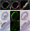
Dual-modality OFDI-NIRF images of atherosclerosis microstructure and inflammatory enzyme activity in a living rabbit. (a) A representative OFDI cross-sectional image. OFDI shows normal artery wall as well as a focal, raised, lipid-containing (L) atherosclerotic lesion at 3–6 o’clock (arrowheads). (b) The OFDI-NIRF fusion image shows a strong NIRF signal in the OFDI-delineated plaque. The NIRF signal reflecting inflammatory protease activity was minimal in the area that appeared to be normal by OFDI. (c) Expanded view of b. 4 o’clock portion (green asterisk) of the plaque demonstrated a stronger NIRF signal than the adjacent regions (blue asterisk) (d) RAM-11 stained section demonstrates a plaque with a dense accumulation of macrophages. (e) Fluorescence microscopy image (20×, stitched) of autofluorescence (green) and protease activity-induced NIR fluorescence (red). Strong NIR fluorescence signal was detected at the luminal surface of the plaque, subtending a similar arc as that of the elevated NIRF signal obtained in vivo. (f) Immunoreactive cathepsin B was detected in the macrophage-rich plaque region. (gi) Expanded views of df, respectively. Macrophage staining by RAM-11 did not appear to vary significantly across the plaque. However, as with NIRF signals in vivo, fluorescence microscopy and IHC for cathepsin B demonstrated heterogeneous protease activity in the plaque with a higher signal at 4 o’clock (green asterisk) compared with that at 5 o’clock (blue asterisk), similar to the pattern observed in the NIR fluorescence microscopic images in h. Scale bars, 500 µm (a,b,df) and 200 µm (c,gi).

References

    1. Lloyd-Jones D, et al. Heart disease and stroke statistics--2009 update: a report from the American Heart Association Statistics Committee and Stroke Statistics Subcommittee. Circulation. 2009;119:480–486. - PubMed
    1. Yun SH, et al. Comprehensive volumetric optical microscopy in vivo. Nat Med. 2006;12:1429–1433. - PMC - PubMed
    1. Huang D, et al. Optical coherence tomography. Science. 1991;254:1178–1181. - PMC - PubMed
    1. Choma M, Sarunic M, Yang C, Izatt J. Sensitivity advantage of swept source and Fourier domain optical coherence tomography. Opt Express. 2003;11:2183–2189. - PubMed
    1. Hausler G, Lindner MW. "Coherence Radar" and "Spectral Radar" - New tools for dermatological diagnosis. Journal of Biomedical Optics. 1998;3:21–31. - PubMed

Publication types