. 2011 Nov 6;17(12):1685-91.
doi: 10.1038/nm.2554.

Cancer cell-selective in vivo near infrared photoimmunotherapy targeting specific membrane molecules

Affiliations

Cancer cell-selective in vivo near infrared photoimmunotherapy targeting specific membrane molecules

Makoto Mitsunaga et al. Nat Med. .

Abstract

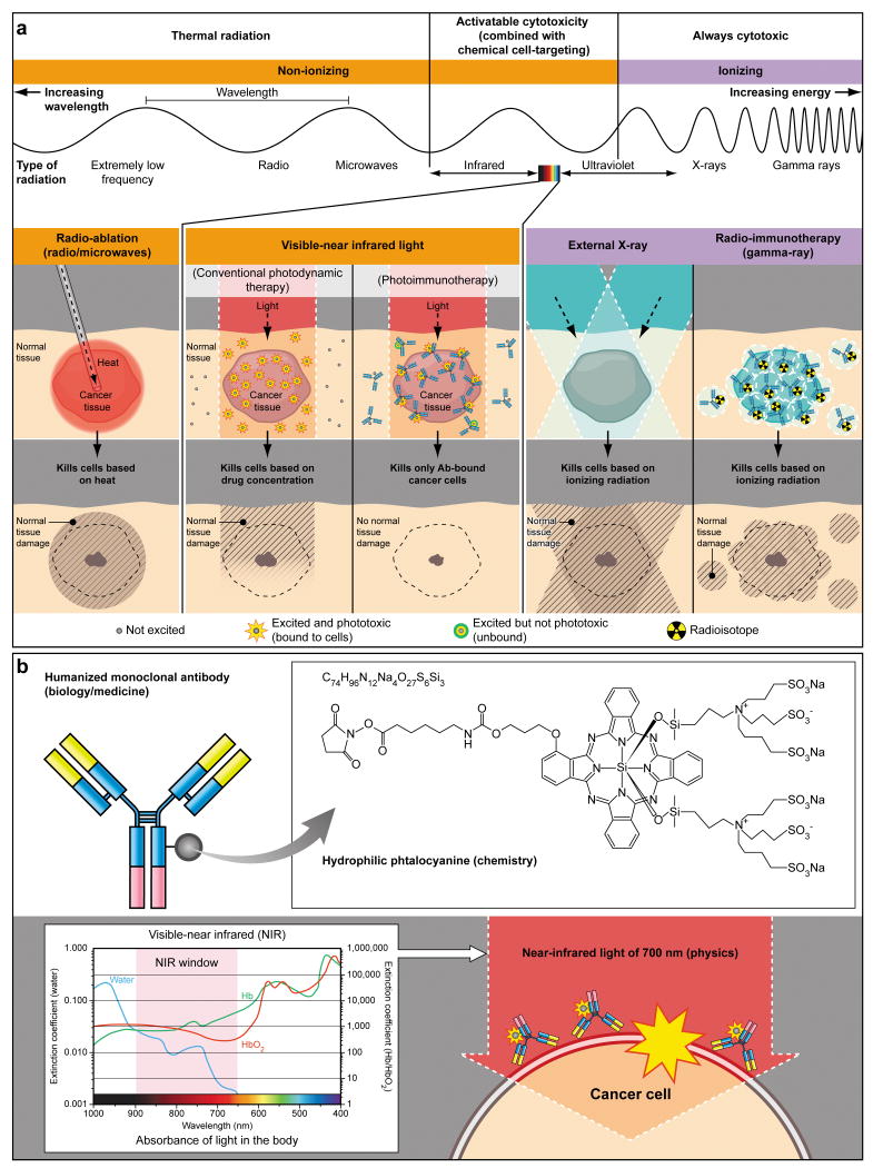
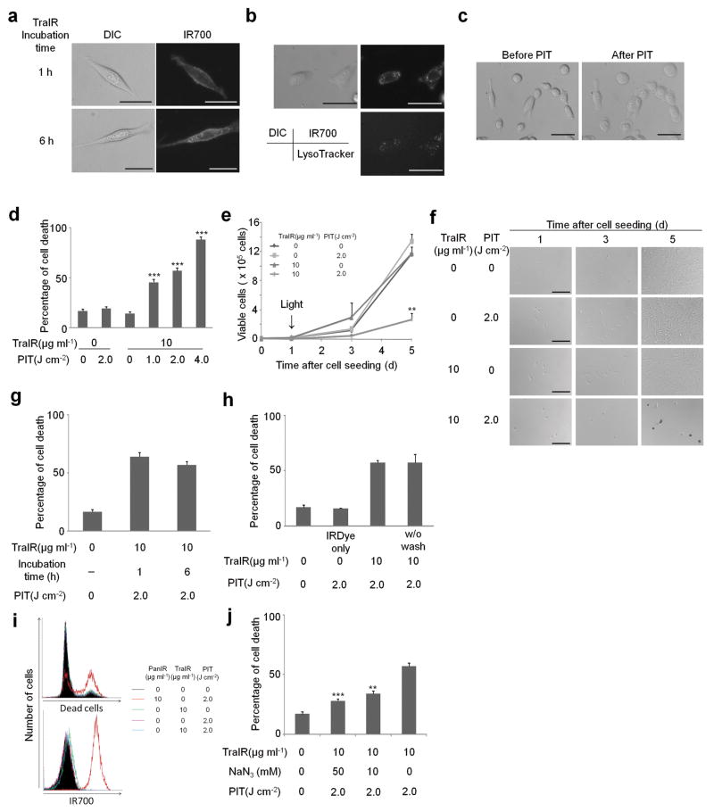
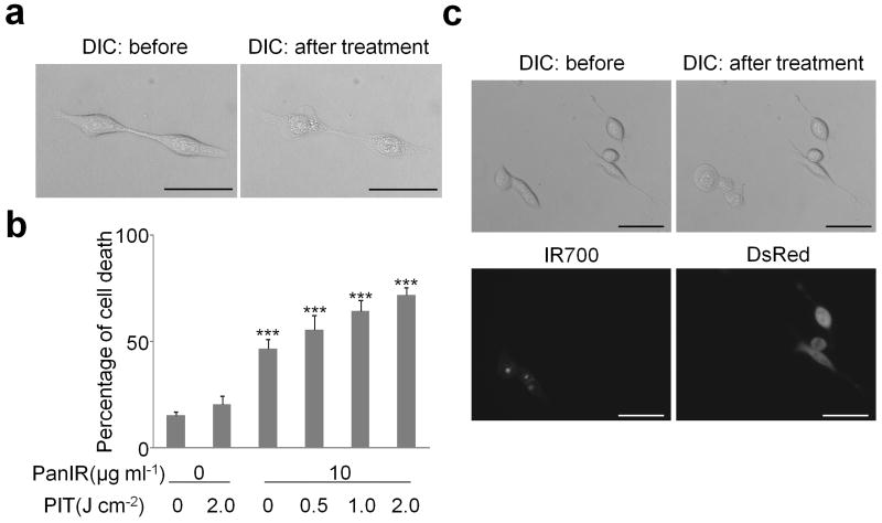
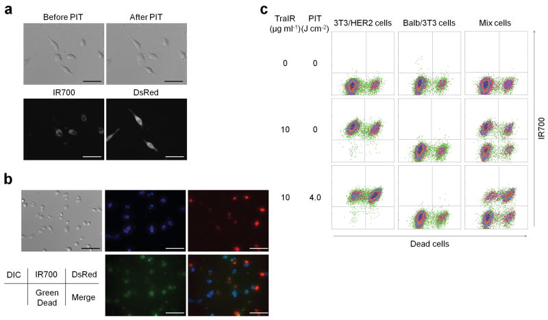
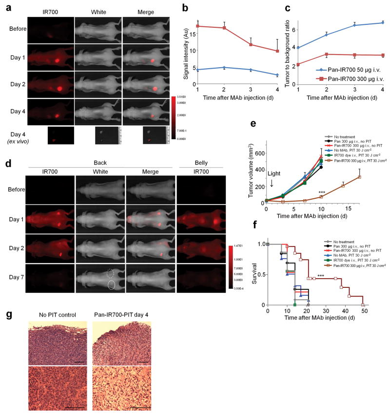
Three major modes of cancer therapy (surgery, radiation and chemotherapy) are the mainstay of modern oncologic therapy. To minimize the side effects of these therapies, molecular-targeted cancer therapies, including armed antibody therapy, have been developed with limited success. In this study, we have developed a new type of molecular-targeted cancer therapy, photoimmunotherapy (PIT), that uses a target-specific photosensitizer based on a near-infrared (NIR) phthalocyanine dye, IR700, conjugated to monoclonal antibodies (mAbs) targeting epidermal growth factor receptors. Cell death was induced immediately after irradiating mAb-IR700-bound target cells with NIR light. We observed in vivo tumor shrinkage after irradiation with NIR light in target cells expressing the epidermal growth factor receptor. The mAb-IR700 conjugates were most effective when bound to the cell membrane and produced no phototoxicity when not bound, suggesting a different mechanism for PIT as compared to conventional photodynamic therapies. Target-selective PIT enables treatment of cancer based on mAb binding to the cell membrane.


Figures

Figure 1
Figure 1
(a) A schema for explaining selective cancer therapy with photoimmunotherapy (PIT) in the context of other physical cancer therapies employing electro-magnetic wave irradiation. Although other physical cancer therapies induce different types of damages in the normal tissue, PIT dedicatedly damages cancer cells without damaging normal cells or tissues. (b) A schema for explaining photo-physical, chemical and biological basis of PIT. Humanized antibodies are employed as a delivary vehicle from the biology and medicine points of view because of its highest binding specificity, greatest in vivo target delivery, low immunogenecity among the clinically applicable targeting reagents. A hydrophilic phtalocyanine is employed as a activatable cytotoxic “Nano-dynamite” reagent from the chemistry points of view because of its great absorption of near infrared light of 700nm and strong cytotoxicity induced only when associating with the cell membrane. Near infrared light of 700nm is employed as an initiator for activating cytotoxicity from the physics points of view because of its high energy among non-harmful non-ionizing photons and great in vivo tissue penetration.
Figure 2
Figure 2. Target specific cell death in response to Tra-IR700 mediated photoimmunotherapy for 3T3/HER2 cells
(a) Different subcellular localization of Tra-IR700 (TraIR). Scale bar, 30 μm. (b) Lysosomal localization of Tra-IR700 6 h after incubation. Scale bar, 50 μm. (c) Microscopic observation of before and after Tra-IR700 mediated photoimmunotherapy (PIT). Scale bar, 50 μm. (d) Irradiation dose dependent and target specific cell death in response to Tra-IR700 mediated PIT. Data are means ± s.e.m. (n = at least 4, *** P < 0.001 vs. non treatment control, Student's t test). (e) Long term growth inhibition in response to Tra-IR700 mediated PIT. Data are means ± s.e.m. (n = 3, ** P < 0.01 vs. non treatment control, Student's t test). (f) Microscopic observation of growth inhibition in response to Tra-IR700 mediated PIT. Scale bar, 100 μm (g) Internalization of Tra-IR700 was not required for phototoxic cell death. Data are means ± s.e.m. (n = 3). (h) Target specific membrane binding of Tra-IR700 only induced phototoxic cell death. Data are means ± s.e.m. (n = 3). (i) HER2 negatively expressing A431 cells did not show phototoxic effects with Tra-IR700 mediated PIT (n = 3). (j) Sodium azide (NaN3) concentration dependent inhibition of phototoxic cell death induced by Tra-IR700 mediated PIT. Data are means ± s.e.m. (n = 3, *** P < 0.001, ** P < 0.01 vs. 2.0 J cm-2 PIT treatment without NaN3 control, Student's t test). DIC: differential interference contrast. PanIR: Pan-IR700.
Figure 3
Figure 3. Target specific cell death in response to Pan-IR700 mediated photoimmunotherapy for EGFR expressing A431 cells
(a) Microscopic observation of before and after Pan-IR700 mediated photoimmunotherapy (PIT). Scale bar, 50 μm. (b) Irradiation dose dependent and target specific cell death in response to Pan-IR700 (PanIR) mediated PIT. Data are means ± s.e.m. (n = at least 4, *** P < 0.001 vs. non treatment control, Student's t test). (c) EGFR expressing cell specific necrotic cell death was induced by Pan-IR700 mediated PIT. Scale bar, 50 μm. DIC: differential interference contrast.
Figure 4
Figure 4. Tra-IR700 mediated photoimmunotherapy for HER2 expressing and non expressing co-cultured cells
(a) Induction of target specific photoimmunotherapy (PIT) lead to HER2 expressing cell specific necrotic cell death. Scale bar, 50 μm. (See also Supplementary Video 2) (b) HER2 specific cell death was confirmed with fluorescence microscopy with LIVE/DEAD Green staining. Scale bar, 100 μm. (c) Flow cytometric analysis for detecting HER2-specific cell death induced by Tra-IR700 (TraIR) mediated PIT. Upper left quadrant: Tra-IR700 positive, live cells; upper right quadrant; Tra-IR700 positive, dead cells; lower left quadrant: Tra-IR700 negative, live cells; lower right quadrant: Tra-IR700 negative, dead cells (n = 3). DIC: differential interference contrast.
Figure 5
Figure 5. Pan-IR700 mediated photoimmunotherapy for HER1 expressing tumors in vivo
(a) HER1 positive A431 tumor (left dorsum) was selectively visualized as early as 1 d after Pan-IR700 injection (50 μg). HER1 negative 3T3/HER2 tumor (right dorsum) did not show detectable fluorescence (n = 5 mice). (b) Fluorescence intensity of IR700 in A431 tumors over time at two different dose of Pan-IR700. Data are means ± s.e.m. (n = 4 each mice). (c) Tumor to background ratio of IR700 fluorescence intensity in A431 tumors over time at two different dose of Pan-IR700. Data are means ± s.e.m. (n = 4 each mice). (d) Biodistribution of Pan-IR700. A431 tumors (both sides of dorsum) were selectively visualized with IR700 fluorescence as early as 1 d after Pan-IR700 injection (300 μg). Right side of the tumor was irradiated with near infrared (NIR) light on day 1, while left side of the tumor was covered with black tape. Tumor shrinkage was confirmed on day 7. Dashed line: irradiated tumor, solid line: non-irradiated tumor. (e) Target specific tumor growth inhibition by Pan-IR700 mediated photoimmunotherapy (PIT) for A431 tumors. PIT was performed on day 1 after Pan-IR700 injection (day 5 after tumor inoculation). Data are means ± s.e.m. (at least n = 12 mice in each group, *** P < 0.001 vs. other control groups, Kruskal–Wallis test with post-test). (f) Kaplan-Meier survival curve analysis of Pan-IR700 mediated PIT for A431 tumors (at least n = 12 mice in each group, *** P < 0.001 vs. other control groups, log-rank test with Bonferroni's correction for multiplicity). (g) Histological observation of treated and non-treated A431 tumors (n = 5 mice, hematoxylin and eosin staining). Scale bar, 100 μm. Pan: panitumumab.

References

    1. Waldmann TA. Immunotherapy: past, present and future. Nat Med. 2003;9:269–277. - PubMed
    1. Reichert JM, Rosensweig CJ, Faden LB, Dewitz MC. Monoclonal antibody successes in the clinic. Nat Biotechnol. 2005;23:1073–1078. - PubMed
    1. Goldenberg DM, Sharkey RM, Paganelli G, Barbet J, Chatal JF. Antibody pretargeting advances cancer radioimmunodetection and radioimmunotherapy. J Clin Oncol. 2006;24:823–834. - PubMed
    1. Pastan I, Hassan R, Fitzgerald DJ, Kreitman RJ. Immunotoxin therapy of cancer. Nat Rev Cancer. 2006;6:559–565. - PubMed
    1. Mew D, Wat CK, Towers GH, Levy JG. Photoimmunotherapy: treatment of animal tumors with tumor-specific monoclonal antibody-hematoporphyrin conjugates. J Immunol. 1983;130:1473–1477. - PubMed

Publication types