Case Reports
. 2012 Feb;123(2):235-45.
doi: 10.1007/s00401-011-0900-5. Epub 2011 Nov 6.

Pathology of immune reconstitution inflammatory syndrome in multiple sclerosis with natalizumab-associated progressive multifocal leukoencephalopathy

Affiliations
Case Reports

Pathology of immune reconstitution inflammatory syndrome in multiple sclerosis with natalizumab-associated progressive multifocal leukoencephalopathy

Imke Metz et al. Acta Neuropathol. 2012 Feb.

Abstract

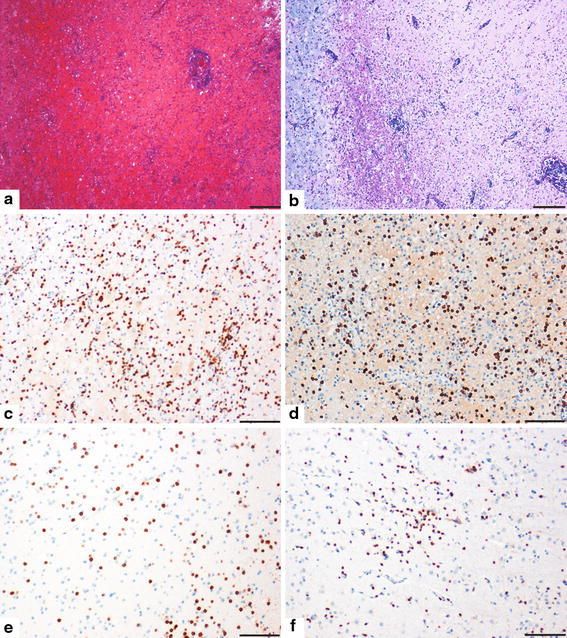
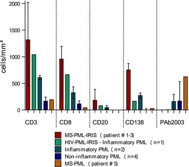
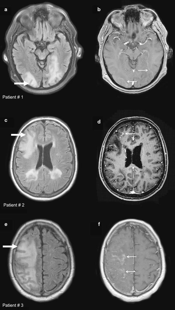
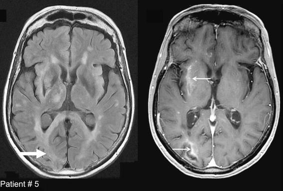
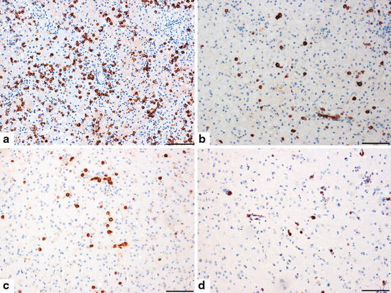
Natalizumab is an approved medication for highly active multiple sclerosis (MS). Progressive multifocal leukoencephalopathy (PML) may occur as a severe side effect of this drug. Here, we describe pathological and radiological characteristics of immune reconstitution inflammatory syndrome (IRIS), which occurs in natalizumab-associated PML after the cessation of therapy, and we differentiate it from ongoing PML. Brain biopsy tissue and MRI scans from five MS patients with natalizumab-associated PML were analyzed and their histology compared with non-MS PML. Histology showed an extensive CD8-dominated T cell infiltrate and numerous macrophages within lesions, and in nondemyelinated white and grey matter, in four out of five cases. Few or no virally infected cells were found. This was indicative of IRIS as known from HIV patients with PML. Outstandingly high numbers of plasma cells were present as compared to non-MS PML and typical MS lesions. MRI was compatible with IRIS, revealing enlarging lesions with a band-like or speckled contrast enhancement either at the lesion edge or within lesions. Only the fifth patient showed typical PML pathology, with low inflammation and high numbers of virally infected cells. This patient showed a similar interval between drug withdrawal and biopsy (3.5 months) to the rest of the cohort (range 2.5-4 months). MRI could not differentiate between PML-associated IRIS and ongoing PML. We describe in detail the histopathology of IRIS in natalizumab-associated PML. PML-IRIS, ongoing PML infection, and MS exacerbation may be impossible to discern clinically alone. MRI may provide some clues for distinguishing different pathologies that can be differentiated histologically. In our individual cases, biopsy helped to clarify diagnoses in natalizumab-associated PML.


Figures

Fig. 1
Fig. 1
Histology of IRIS is characterized by extensive T-cell inflammation in natalizumab-associated PML. An inflammatory demyelinating lesion with pronounced inflammation is shown (a H&E, b LFB/PAS). T cells are dominated by CD8+ T cells (c CD3, d CD8). Inflammation is also evident in adjacent nondemyelinated white and grey matter (e + f CD3). Original magnifications: a + b ×40; cf ×100. Scale bars 200 μm. Patient 2
Fig. 2
Fig. 2
Quantification of inflammation and virally infected cells within demyelinating lesions in MS–PML–IRIS, ongoing MS–PML, and non-MS PML. Numbers of T cells (CD3), CD8+ T cells (CD8), B cells (CD20), plasma cells (CD138), and virally infected cells (PAb2003) were analyzed within demyelinating lesions in MS–PML–IRIS, ongoing MS–PML, and control groups. The median and range are indicated
Fig. 3
Fig. 3
MRI characteristics of patients with natalizumab-associated PML and histologically confirmed IRIS. Axial FLAIR images (a, c, e) of MS patients, cases 1, 2, and 3, with corresponding contrast-enhanced T1-weighted images (b, d, f) are shown. Different patterns of enhancement in IRIS can be seen, ranging from faint peripheral enhancement to intense enhancement of the lesions (open arrows in b, d, and f). The area of biopsy is indicated (closed arrows in a, c, and e)
Fig. 4
Fig. 4
MRI characteristics of a patient with histologically confirmed active natalizumab-associated PML. Axial FLAIR (a) and corresponding contrast-enhanced T1-weighted images (b) are shown (case 5). Linear enhancement at the lesion edge as well as diffuse and speckled enhancement within lesions are found (open arrows in b). The enhancement pattern does not allow differentiation between active PML and IRIS. The biopsied area is indicated (closed arrow in a)
Fig. 5
Fig. 5
High numbers of plasma cells are evident in natalizumab-associated PML with IRIS. Strikingly high numbers of plasma cells are found in MS–PML–IRIS lesions (a). Lower numbers are present in inflammatory PML cases (b). In MS–PML–IRIS, plasma cells are also evident in nondemyelinated white and grey matter (c, d) (ad CD138). Original magnifications: ad ×100. Scale bars 200 μm. a + d Patient 2, b inflammatory PML control, c patient 1

Similar articles

Cited by

References

    1. Berger JR. Progressive multifocal leucoencephalopathy and newer biological agents. Drug Saf. 2010;33:969–983. doi: 10.2165/11537510-000000000-00000. - DOI - PubMed
    1. Clifford DB, De LA, Simpson DM, Arendt G, Giovannoni G, Nath A. Natalizumab-associated progressive multifocal leukoencephalopathy in patients with multiple sclerosis: lessons from 28 cases. Lancet Neurol. 2010;9:438–446. doi: 10.1016/S1474-4422(10)70028-4. - DOI - PubMed
    1. Du Pasquier RA, Kuroda MJ, Zheng Y, Jean-Jacques J, Letvin NL, Koralnik IJ. A prospective study demonstrates an association between JC virus-specific cytotoxic T lymphocytes and the early control of progressive multifocal leukoencephalopathy. Brain. 2004;127:1970–1978. doi: 10.1093/brain/awh215. - DOI - PubMed
    1. Engelhardt B, Kappos L. Natalizumab: targeting alpha4-integrins in multiple sclerosis. Neurodegener Dis. 2008;5:16–22. doi: 10.1159/000109933. - DOI - PubMed
    1. Gasnault J, Kahraman M, de Herve MG, Durali D, Delfraissy JF, Taoufik Y. Critical role of JC virus-specific CD4 T-cell responses in preventing progressive multifocal leukoencephalopathy. AIDS. 2003;17:1443–1449. doi: 10.1097/00002030-200307040-00004. - DOI - PubMed

Publication types

MeSH terms

Substances