Review
. 2012 Mar;34(2):261-80.
doi: 10.1007/s00281-011-0292-6. Epub 2011 Nov 6.

Innate and adaptive immune responses against Staphylococcus aureus skin infections

Affiliations
Review

Innate and adaptive immune responses against Staphylococcus aureus skin infections

Sheila Krishna et al. Semin Immunopathol. 2012 Mar.

Abstract

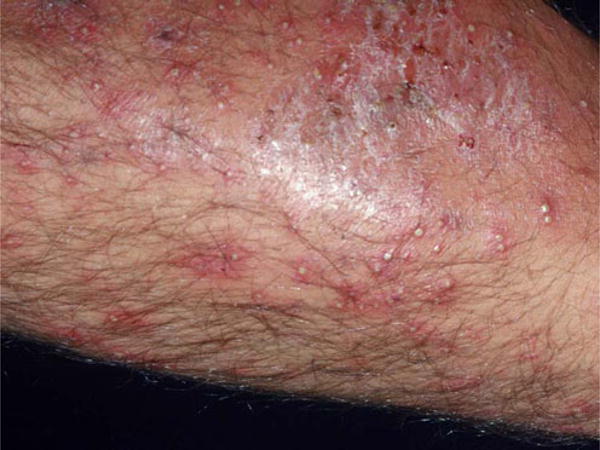
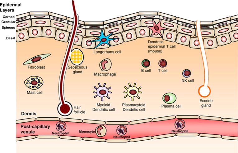
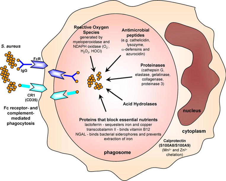
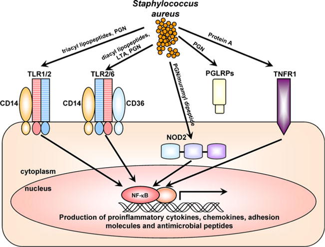
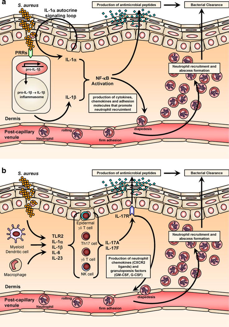
Staphylococcus aureus is an important human pathogen that is responsible for the vast majority of bacterial skin and soft tissue infections in humans. S. aureus can also become more invasive and cause life-threatening infections such as bacteremia, pneumonia, abscesses of various organs, meningitis, osteomyelitis, endocarditis, and sepsis. These infections represent a major public health threat due to the enormous numbers of these infections and the widespread emergence of methicillin-resistant S. aureus (MRSA) strains. MSRA is endemic in hospitals worldwide and is rapidly spreading throughout the normal human population in the community. The increasing frequency of MRSA infections has complicated treatment as these strains are more virulent and are increasingly becoming resistant to multiple different classes of antibiotics. The important role of the immune response against S. aureus infections cannot be overemphasized as humans with certain genetic and acquired immunodeficiency disorders are at an increased risk for infection. Understanding the cutaneous immune responses against S. aureus is essential as most of these infections occur or originate from a site of infection or colonization of the skin and mucosa. This review will summarize the innate immune responses against S. aureus skin infections, including antimicrobial peptides that have direct antimicrobial activity against S. aureus as well as pattern recognition receptors and proinflammatory cytokines that promote neutrophil abscess formation in the skin, which is required for bacterial clearance. Finally, we will discuss the recent discoveries involving IL-17-mediated responses, which provide a key link between cutaneous innate and adaptive immune responses against S. aureus skin infections.


Figures

Fig. 1
Fig. 1
S. aureus folliculitis. Numerous infected hair follicles present as follicularly based erythematous, warm, edematous papules and pustules on this extremity (Courtesy of the Victor D. Newcomer collection at UCLA and Logical Images, Inc.)
Fig. 2
Fig. 2
The physical and immune barrier of the skin. The epidermis is composed of layers of keratinocytes, including the corneal, granular, spinous, and basal layers. There are sweat glands, sebaceous glands, and hair follicles that span these layers. In addition, there are many resident immune cells in skin that participate in immune responses. In the epidermidis, there are Langerhans cells and in mouse epidermis there are DETCs. In the dermis, there are mast cells, macrophages, myeloid and plasmacytoid dendritic cells, B and T cells, NK cells, and plasma cells. Neutrophils, monocytes, and other immune cells are recruited from the circulation also participate in cutaneous immune responses
Fig. 3
Fig. 3
Neutrophil antimicrobial mechanisms against S. aureus. Neutrophils engulf opsonized bacteria using Fc and complement receptors into phagosomes. Inside phagosomes, neutrophils possess multiple distinct mechanisms to promote bacterial clearance. Oxidative burst generates reactive oxygen species (such as O2, H2O2, and HOCl) through myeloperoxidase and NADPH oxidase, which can directly kill bacteria but also produce a charge in the phagosome membrane to induce enzymatic killing. Antimicrobial peptides such as cathelicidin (LL-37), lysozyme, α-defensins, and azurocidin also have direct antimicrobial activity. Proteinases such as cathepsin G, neutrophil elastase, gelatinase, collagenase and proteinase 3, and acid hydrolases degrade bacterial components. Neutrophils also express proteins that sequester essential nutrients from bacteria to limit their growth such as lactoferrin (which sequesters iron and copper), transcobalamin II (which binds vitamin B12), and neutrophil gelatinase associated lipocalin (NGAL; which binds bacterial siderophores, preventing the extraction of iron). If S. aureus enters the cytoplasm of neutrophils, there is an abundance of calprotectin, which sequesters Mn2+ and Zn2+ to inhibit bacterial growth
Fig. 4
Fig. 4
Pattern recognition receptors (PRRs) that recognize S. aureus. PRRs involved in recognizing components of S. aureus and cellular localization of these PRRs are shown. Toll-like receptor 2 (TLR2) heterodimerizes with TLR1 and TLR6 to recognize tri- and di-acyl lipopeptides, respectively (including S. aureus lipoteichoic acid [LTA], which is diacylated). TLR2 also recognizes S. aureus peptidoglycan (PGN) and CD14 and CD36 act as TLR2 co-receptors. Nucleotide-binding oligomerization domain containing 2 (NOD2) is an intracellular cytoplasmic receptor that recognizes the S. aureus peptidoglycan breakdown product muramyl dipeptide. Peptidoglycan receptor proteins (PGRPs or PGLYRPs) are secreted proteins that recognize S. aureus PGN. TNFR1 is a cell surface receptor that is activated by TNF-α but has also been shown to recognize S. aureus protein A. In general, signaling from these PRRs promotes activation of NF-κB and other transcription factors that induce transcription of proinflammatory cytokines, chemokines, adhesion molecules, and antimicrobial peptides that are involved in cutaneous host defense against S. aureus
Fig. 5
Fig. 5
Cytokine responses that promote clearance of S. aureus skin infections. a IL-1 response against S. aureus. In response to S. aureus, IL-1α is produced and released by keratinocytes in an autocrine signaling loop. In addition, S. aureus skin infection results in activation of PRRs and the inflammasome by resident and recruited cells (such as macrophages and dendritic cells), which leads to production and secretion of active IL-1β. b IL-17 mediated response against S. aureus. IL-17A and IL-17F are produced by multiple different types of cells in the skin, including Th17 cells, γδ T cells, and NK cells (and perhaps mast cells and neutrophils) in response to different signals such as activation by TLR2, IL-1α/β, IL-6, and IL-23. IL-17A and IL-17F mediate immune responses by binding to the IL-17R, which is expressed primarily by keratinocytes. Both the IL-1 and IL-17 responses promote neutrophil recruitment and abscess formation and keratinocyte production of antimicrobial peptides (e.g., hBD2, hBD3, and LL-37) to help control S. aureus skin infections and mediate bacterial clearance

References

    1. McCaig LF, McDonald LC, Mandal S, Jernigan DB. Staphylococcus aureus-associated skin and soft tissue infections in ambulatory care. Emerg Infect Dis. 2006;12:1715–1723. - PMC - PubMed
    1. Moran GJ, Krishnadasan A, Gorwitz RJ, Fosheim GE, McDougal LK, Carey RB, Talan DA. Methicillin-resistant S. aureus infections among patients in the emergency department. N Engl J Med. 2006;355:666–674. - PubMed
    1. Klevens RM, Morrison MA, Nadle J, Petit S, Gershman K, Ray S, Harrison LH, Lynfield R, Dumyati G, Townes JM, Craig AS, Zell ER, Fosheim GE, McDougal LK, Carey RB, Fridkin SK. Invasive methicillin-resistant Staphylococcus aureus infections in the United States. JAMA. 2007;298:1763–1771. - PubMed
    1. Daum RS. Clinical practice. Skin and soft-tissue infections caused by methicillin-resistant Staphylococcus aureus. N Engl J Med. 2007;357:380–390. - PubMed
    1. Deleo FR, Otto M, Kreiswirth BN, Chambers HF. Community-associated methicillin-resistant Staphylococcus aureus. Lancet. 2010;375:1557–1568. - PMC - PubMed

Publication types

MeSH terms

LinkOut - more resources