. 2011 Nov;32(5):437-44.
doi: 10.1007/s10059-011-0107-6. Epub 2011 Nov 1.

Simvastatin promotes osteogenic differentiation of mouse embryonic stem cells via canonical Wnt/β-catenin signaling

Affiliations

Simvastatin promotes osteogenic differentiation of mouse embryonic stem cells via canonical Wnt/β-catenin signaling

Ling Juan Qiao et al. Mol Cells. 2011 Nov.

Abstract

Simvastatin, an inhibitor of 3-hydroxy-3-methylglutaryl coenzyme A (HMG-CoA) reductase, has been known to reduce cholesterol biosynthesis. However, recent studies demonstrate that simvastatin shows diverse cholesterol-independent functions including cellular differentiation. In this study, we investigated the stimulatory effect of simvastatin on the osteogenic differentiation of mouse embryonic stem cells (ESCs). The osteogenic effect of simvastatin was observed at relatively low doses (ranging from 1 nM to 200 nM). Incubation of ESCs in simvastatin-supplemented osteogenic medium significantly increased alkaline phosphatase (ALP) activity at day 7. The matrix mineralization was also augmented and demonstrated pivotal levels after 14 days incubation of simvastatin. Osteogenic differentiation of ESCs by simvastatin was determined by upregulation of the mRNA expression of runtrelated gene 2 (Runx2), osterix (OSX), and osteocalcin (OCN) as osteogenic transcription factors. Moreover, the increased protein expression of OCN, osteopontin (OPN), and collagen type I (Coll I) was assessed using Western blot analysis and immunocytochemistry. However, the blockage of canonical Wnt signaling by DKK-1 downregulated simvastatin-induced ALP activity and the mRNA expression of each osteogenic transcription factor. Furthermore, the β-catenin specific siRNA transfection decreased the protein levels of OCN, OPN, and Coll I. Collectively, these findings suggest that simvastatin enhances the differentiation of ESCs toward osteogenic lineage through activation of canonical Wnt/β-catenin signaling.


Figures
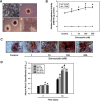
Fig. 1.
Fig. 1.. Effect of simvastatin on osteogenic differentiation of ESCs. (A) Morphology of the cells under a light microscope. Undifferentiated ES cells (a), suspended EBs (b), EBs incubated in osteogenic medium for 1 day (c), and EBs cultured in osteogenic medium with simvastatin for 7 days (d) (magnification 20×). Cells were incubated in osteogenic medium with simvastatin (1, 10, 100, 200 nM) for 4, 7, and 14 days each, then (B) ALP activity or (C) Alizarin red staining was assessed as described in “Materials and Methods”. Each microscopic image shown is a representative of five separate experi-ments. The size bars on panel A represent 50 μm. (D) ARS quanti-fication was assessed on days 7 and 14 as described in “Materials and Methods”. The values reported are the mean ± S.D. of five independent experiments. *P < 0.05 or # P < 0.001 vs. control value.
Fig. 2.
Fig. 2.. Effects of different concentrations of simvastatin on the mRNA expression of Runx2, OSX, and OCN. The mRNA levels of (A) Runx2, (B) OSX, and (C) OCN were analyzed using the real time RT-PCR technique after a 4 day-osteogenic induction. The values reported are the mean ± S.D. of three independent experiments. *P < 0.05 vs. control value.
Fig. 3.
Fig. 3.. Effect of simvastatin on OCN, OPN, and collagen type I protein levels. (A) Cells were treated with simvastatin (1, 10, 100, 200 nM) for 7 days and then the protein levels of OCN, OPN, and collagen type I were determined by Western blot analysis using total protein lysates. (B) The panels (bars) denote the mean ± S.D. of three experiments for each condition determined from densitometry relative to β-actin. *P < 0.05 vs. control value.
Fig. 4.
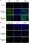
Fig. 4.. Immunofluorescence staining with antibodies of OPN and collagen type I. Cells were incubated in the presence of 100 or 200 nM simvastatin for 7 days then (A) OPN and (B) collagen type I were detected by the immunostaining method. The nuclei were stained with DAPI as shown in blue. A representative result from the four independent experiments is shown.
Fig. 5.
Fig. 5.. Inhibitory effect of DKK-1 on simvastatin-induced osteogenic differentiation of ESCs. Cells were pretreated with 0.5 μg/ml DKK-1 1 h before the addition of simvastatin (100 nM) and (A) processed for the analysis of ALP activity after 7 days of incubation. (B) mRNA levels of Runx2, OSX, and OCN were also measured by real time RT-PCR. The results shown are the mean ± S.D. from five separate experiments. # P < 0.001 vs. the untreated control values, ## P < 0.05 and ### P < 0.001 vs. simvastatin treatment alone.
Fig. 6.
Fig. 6.. Effect of knockdown of β-catenin on simvastatin-stimulated osteogenesis of ESCs. Cells were transfected with either β-catenin- or GFP-specific siRNA using LipofectAMINE 2000 and after 24 h of transfection, and the cells were then treated with simvastatin (100 nM). (A) The protein levels of β-catenin according to the siRNA transfection were determined by Western blotting after 2 days of transfection. (B) The protein levels of OCN, OPN, and collagen type I were also detected after 7 days of incubation. A representative result from the four independent experiments is shown.

References

    1. Baek K.H., Lee W.Y., Oh K.W., Tae H.J., Lee J.M., Lee E.J., Han J.H., Kang M.I., Cha B.Y., Lee K.W., et al. The effect of simvastatin on the proliferation and differentiation of human bone marrow stromal cells. J. Korean Med. Sci. (2005);20:438–444. - PMC - PubMed
    1. Bielby R.C., Boccaccini A.R., Polak J.M., Buttery L.D. In vitro differentiation and in vivo mineralization of osteogenic cells derived from human embryonic stem cells. Tissue Eng. (2004);10:1518–1525. - PubMed
    1. Bradford M.M. A rapid and sensitive method for the quantitation of microgram quantities of protein utilizing the principle of protein-dye binding. Anal. Biochem. (1976);72:248–254. - PubMed
    1. Bratt-Leal A.M., Carpenedo R.L., McDevitt T.C. Engineering the embryoid body microenvironment to direct embryonic stem cell differentiation. Biotechnol. Prog. (2009);25:43–51. - PMC - PubMed
    1. Buttery L.D., Bourne S., Xynos J.D., Wood H., Hughes F.J., Hughes S.P., Episkopou V., Polak J.M. Differentiation of osteoblasts and in vitro bone formation from murine embryonic stem cells. Tissue Eng. (2001);7:89–99. - PubMed

Publication types

MeSH terms