Review
. 2012 Mar 1;302(5):H1031-49.
doi: 10.1152/ajpheart.00899.2011. Epub 2011 Nov 4.

How NaCl raises blood pressure: a new paradigm for the pathogenesis of salt-dependent hypertension

Affiliations
Review

How NaCl raises blood pressure: a new paradigm for the pathogenesis of salt-dependent hypertension

Mordecai P Blaustein et al. Am J Physiol Heart Circ Physiol. .

Abstract

Excess dietary salt is a major cause of hypertension. Nevertheless, the specific mechanisms by which salt increases arterial constriction and peripheral vascular resistance, and thereby raises blood pressure (BP), are poorly understood. Here we summarize recent evidence that defines specific molecular links between Na(+) and the elevated vascular resistance that directly produces high BP. In this new paradigm, high dietary salt raises cerebrospinal fluid [Na(+)]. This leads, via the Na(+)-sensing circumventricular organs of the brain, to increased sympathetic nerve activity (SNA), a major trigger of vasoconstriction. Plasma levels of endogenous ouabain (EO), the Na(+) pump ligand, also become elevated. Remarkably, high cerebrospinal fluid [Na(+)]-evoked, locally secreted (hypothalamic) EO participates in a pathway that mediates the sustained increase in SNA. This hypothalamic signaling chain includes aldosterone, epithelial Na(+) channels, EO, ouabain-sensitive α(2) Na(+) pumps, and angiotensin II (ANG II). The EO increases (e.g.) hypothalamic ANG-II type-1 receptor and NADPH oxidase and decreases neuronal nitric oxide synthase protein expression. The aldosterone-epithelial Na(+) channel-EO-α(2) Na(+) pump-ANG-II pathway modulates the activity of brain cardiovascular control centers that regulate the BP set point and induce sustained changes in SNA. In the periphery, the EO secreted by the adrenal cortex directly enhances vasoconstriction via an EO-α(2) Na(+) pump-Na(+)/Ca(2+) exchanger-Ca(2+) signaling pathway. Circulating EO also activates an EO-α(2) Na(+) pump-Src kinase signaling cascade. This increases the expression of the Na(+)/Ca(2+) exchanger-transient receptor potential cation channel Ca(2+) signaling pathway in arterial smooth muscle but decreases the expression of endothelial vasodilator mechanisms. Additionally, EO is a growth factor and may directly participate in the arterial structural remodeling and lumen narrowing that is frequently observed in established hypertension. These several central and peripheral mechanisms are coordinated, in part by EO, to effect and maintain the salt-induced elevation of BP.


Figures

Fig. 1.
Fig. 1.
Overview of the proposed pathways by which salt and endogenous ouabain (EO) secretion effect increased central sympathoexcitation, enhanced peripheral sympathetic nerve activity, and augmented arterial constriction in essential hypertension. As illustrated here, the initiating factor is high dietary salt (box on the left). ACTH, adrenocorticotropic hormone; CSF, cerebrospinal fluid.
Fig. 2.
Fig. 2.
Prolonged administration of ouabain, but not digoxin, to normal rats elevates mean blood pressure (MBP) with a delay of 1 to 2 wk. Digitoxin also does not elevate blood pressure (not shown). Digoxin and digitoxin both normalize MBP in the ouabain-treated hypertensive rats. Blood pressure was measured by tail-cuff plethysmography. Group 1, phosphate-buffered saline (vehicle) delivered by Alzet osmotic minipump (orange circles; n = 8); group 2, digoxin (30 μg·kg−1·day−1) delivered by minipump (blue squares; n = 8); group 3, ouabain (15 μg·kg−1·day−1) delivered by minipump (green inverted triangles; n = 24). At 35 days, group 3 was divided into 3 subgroups (n = 8 each). All 3 continued to receive ouabain; in addition, all 3 received a second minipump implant that delivered vehicle (group 3A, green inverted triangles), digoxin (30 μg·kg−1·day−1; and group 3B, red circles) or digitoxin (30 μg·kg−1·day−1; group 3C, black circles). *P < 0.05 vs. group 3A; ***P < 0.001 vs. ouabain (group 3); #P < 0.0005 vs. vehicle (group 1); **P < 0.001 vs. digoxin (group 2). Data from Manuta et al. (171), which contains full details.
Fig. 3.
Fig. 3.
Diagram of the proposed hypothalamic Na+-aldosterone-EO-angiotensin II (ANG II) neuromodulatory pathway by which elevated CSF [Na+] increases central sympathoexcitatory pathways. AT1R, ANG II type-1 receptor; ENaC, benzamil-sensitive epithelial Na+ channel; MR, mineralocorticoid receptor; OVLT, organum vasculosum of the lamina terminalis; PVN, paraventricular nucleus; SON, supraoptic nucleus; SFO, subfornical organ. The PVN and SON are hypothalamic nuclei; the OVLT and SFO are circumventricular organs that, together with the median preoptic nucleus of the hypothalamus, are located anteroventral to the third ventricle, in the so-called AV3V region. Revised from Leenen (148).
Fig. 4.
Fig. 4.
Diagram of ouabain/EO-regulated α2 Na+ pump-modulated Ca2+ signaling at the plasma membrane (PM)-sarco(endo)plasmic reticulum junction (PLasmERosome). EO reduces Na+ extrusion by the α2 Na+ pump, thereby increasing local [Na+] in the junctional space (JS) and reducing Ca2+ extrusion by the Na/Ca exchanger (NCX). This enhances Ca2+ signaling and, in arterial smooth muscle, contraction. PMCA, PM Ca2+ pump; ATP, adenosine triphosphate; ADP, adenosine diphosphate; Pi, inorganic phosphate; GPCR, G protein-coupled receptor; AR, agonist receptor; GP, G protein; PLC, phospholipase C; IP3, inositol 1,4,5-trisphosphate; IP3R, IP3, receptor; DAG, diacylglycerol; ROC, receptor-operated channel, and SOC, store-operated channel [both composed of transient receptor potential cation channel (TRPC) proteins]; SERCA, sarco(endo)plasmic reticulum Ca2+ ATPase pump; ECF, extracellular fluid; SR, sarcoplasmic reticulum; RyR, ryanodine receptor. Revised from Blaustein and Wier (25).
Fig. 5.
Fig. 5.
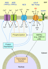
Diagram of ouabain/EO-activated, α2 Na+ pump/Src-mediated signaling cascade (the EO/α2 Na+ pump signalosome). EO binding to the α2 Na+ pump activates a Src (Src family tyrosine kinase), which, in turn, activates mitogen-activated protein kinases (MAPKs) such as P38 and extracellular signal-regulated kinases (ERK1/2). This leads to altered expression and/or phosphorylation of several ion transport proteins including the α2 Na+ pump, NCX, and ROC and SOC. As described in main text, these mechanisms are cell type specific. Ouabain/EO triggers upregulation of Ca2+ signaling proteins and vasoconstriction in arterial myocytes and attenuated vasodilation in endothelial cells. This slow signaling pathway thus modulates the rapid Ca2+ signaling mechanisms diagrammed in Fig. 4. EGFR, epidermal growth factor receptor.

References

    1. Abarquez RF., Jr Digitalis in the treatment of hypertension. A prelminary report. Acta Med Philipp 3: 161–170, 1967 - PubMed
    1. Abboud FM. The sympathetic system in hypertension. State-of-the-art review. Hypertension 4: 208–225, 1982 - PubMed
    1. Abrams JM, Engeland WC, Osborn JW. Effect of intracerebroventricular benzamil on cardiovascular and central autonomic responses to DOCA-salt treatment. Am J Physiol Regul Integr Comp Physiol 299: R1500–R1510, 2010 - PMC - PubMed
    1. Aileru AA, De Albuquerque A, Hamlyn JM, Manunta P, Shah JR, Hamilton MJ, Weinreich D. Synaptic plasticity in sympathetic ganglia from acquired and inherited forms of ouabain-dependent hypertension. Am J Physiol Regul Integr Comp Physiol 281: R635–R644, 2001 - PubMed
    1. Amin MS, Wang HW, Reza E, Whitman SC, Tuana BS, Leenen FH. Distribution of epithelial sodium channels and mineralocorticoid receptors in cardiovascular regulatory centers in rat brain. Am J Physiol Regul Integr Comp Physiol 289: R1787–R1797, 2005 - PubMed

Publication types