

AMD3100 disrupts the cross-talk between chronic lymphocytic leukemia cells and a mesenchymal stromal or nurse-like cell-based microenvironment: pre-clinical evidence for its association with chronic lymphocytic leukemia treatments
- PMID: 22058221
- PMCID: PMC3347654
- DOI: 10.3324/haematol.2011.052779
AMD3100 disrupts the cross-talk between chronic lymphocytic leukemia cells and a mesenchymal stromal or nurse-like cell-based microenvironment: pre-clinical evidence for its association with chronic lymphocytic leukemia treatments
Abstract
Background: Interactions with the microenvironment, such as bone marrow mesenchymal stromal cells and nurse-like cells, protect chronic lymphocytic leukemia cells from spontaneous and drug-induced apoptosis. This protection is partially mediated by the chemokine SDF-1α (CXCL12) and its receptor CXCR4 (CD184) present on the chronic lymphocytic leukemia cell surface.
Design and methods: Here, we investigated the ability of AMD3100, a CXCR4 antagonist, to sensitize chronic lymphocytic leukemia cells to chemotherapy in a chronic lymphocytic leukemia/mesenchymal stromal cell based or nurse-like cell based microenvironment co-culture model.
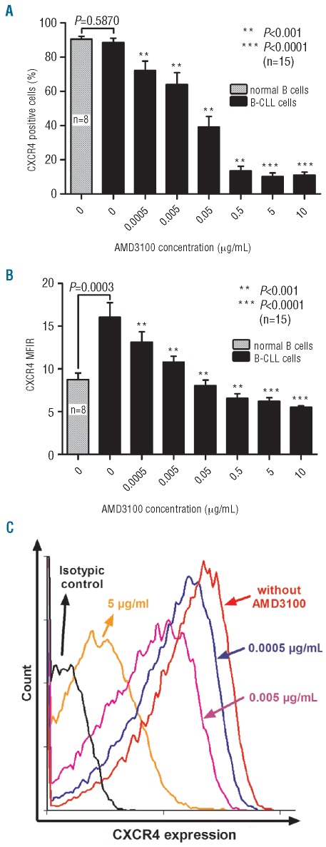
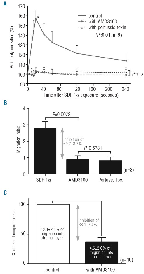
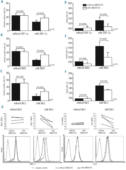
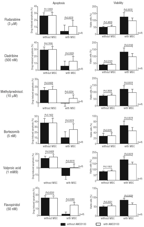
Results: AMD3100 decreased CXCR4 expression signal (n=15, P=0.0078) and inhibited actin polymerization/migration in response to SDF-1α (n=8, P<0.01) and pseudoemperipolesis (n=10, P=0.0010), suggesting that AMD3100 interferes with chronic lymphocytic leukemia cell trafficking. AMD3100 did not have a direct effect on apoptosis when chronic lymphocytic leukemia cells were cultured alone (n=10, P=0.8812). However, when they were cultured with SDF-1α, mesenchymal stromal cells or nurse-like cells (protecting them from apoptosis, P<0.001), chronic lymphocytic leukemia cell pre-treatment with AMD3100 significantly inhibited these protective effects (n=8, P<0.01) and decreased the expression of the anti-apoptotic proteins MCL-1 and FLIP. Furthermore, combining AMD3100 with various drugs (fludarabine, cladribine, valproïc acid, bortezomib, flavopiridol, methylprednisolone) in our mesenchymal stromal cell co-culture model enhanced drug-induced apoptosis (n=8, P<0.05) indicating that AMD3100 could mobilize chronic lymphocytic leukemia cells away from their protective microenvironment, making them more accessible to conventional therapies.
Conclusions: Taken together, these data demonstrate that interfering with the SDF-1α/CXCR4 axis by using AMD3100 inhibited chronic lymphocytic leukemia cell trafficking and microenvironment-mediated protective effects. Combining AMD3100 with other drugs may, therefore, represent a promising therapeutic approach to kill chronic lymphocytic leukemia cells.
Figures
References
-
- Defoiche J, Debacq C, Asquith B, Zhang Y, Burny A, Bron D, et al. Reduction of B cell turnover in chronic lymphocytic leukaemia. Br J Haematol. 2008;143(2):240–7. - PubMed
-
- Dighiero G, Travade P, Chevret S, Fenaux P, Chastang C, Binet JL. B-cell chronic lymphocytic leukemia: present status and future directions. French Cooperative Group on CLL. Blood. 1991;78(8):1901–14. - PubMed
-
- Collins RJ, Verschuer LA, Harmon BV, Prentice RL, Pope JH, Kerr JF. Spontaneous programmed death (apoptosis) of B-chronic lymphocytic leukaemia cells following their culture in vitro. Br J Haematol. 1989;71(3):343–50. - PubMed
-
- Lagneaux L, Delforge A, De Bruyn C, Bernier M, Bron D. Adhesion to bone marrow stroma inhibits apoptosis of chronic lymphocytic leukemia cells. Leuk Lymphoma. 1999;35(5–6):445–53. - PubMed
-
- Caligaris-Cappio F. Role of the microenvironment in chronic lymphocytic leukaemia. Br J Haematol. 2003;123(3):380–8. - PubMed
Publication types
MeSH terms
Substances
LinkOut - more resources
Full Text Sources
