. 2011 Aug 1:41:99-132.
doi: 10.1146/annurev-matsci-062910-100429.

Mussel-Inspired Adhesives and Coatings

Affiliations

Mussel-Inspired Adhesives and Coatings

Bruce P Lee et al. Annu Rev Mater Res. .

Abstract

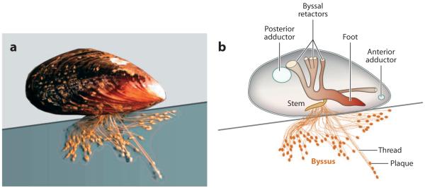
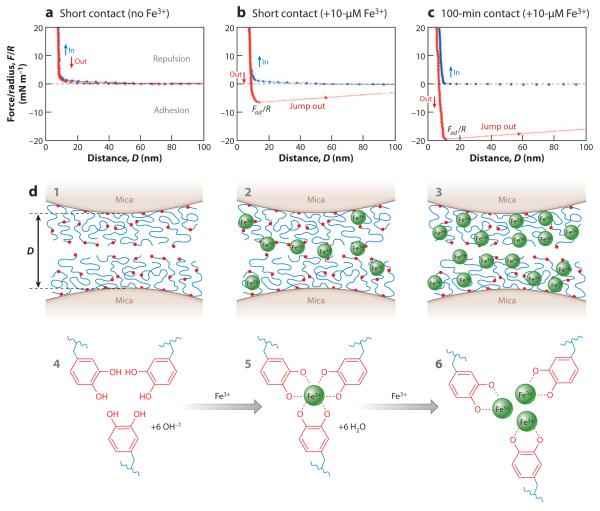
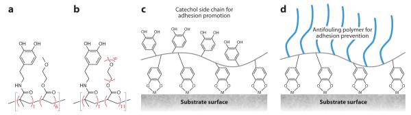
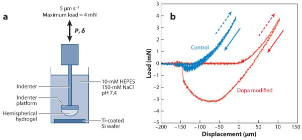
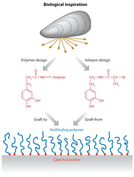
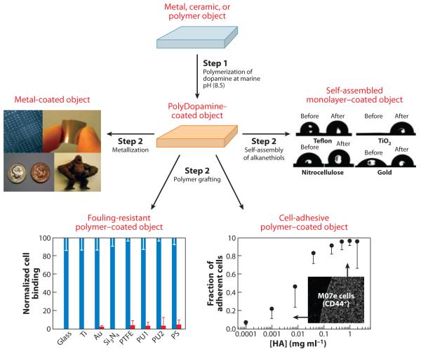
Mussels attach to solid surfaces in the sea. Their adhesion must be rapid, strong, and tough, or else they will be dislodged and dashed to pieces by the next incoming wave. Given the dearth of synthetic adhesives for wet polar surfaces, much effort has been directed to characterizing and mimicking essential features of the adhesive chemistry practiced by mussels. Studies of these organisms have uncovered important adaptive strategies that help to circumvent the high dielectric and solvation properties of water that typically frustrate adhesion. In a chemical vein, the adhesive proteins of mussels are heavily decorated with Dopa, a catecholic functionality. Various synthetic polymers have been functionalized with catechols to provide diverse adhesive, sealant, coating, and anchoring properties, particularly for critical biomedical applications.


Figures

Figure 1
Figure 1
The influence of water on the work of adhesion (WA) between an epoxy adhesive and an aluminum surface. WA is derived from the summation of surface energy products due to dispersion interactions (γ1dγ2d) and those due to polar interactions (γ1pγ2p) under clean-room (left) and wet (right) conditions. From data in Pocius (5).
Figure 2
Figure 2
Adhesion in the marine mussel Mytilus californianus. (a) Adult mussel (5 cm length) displaying an extensive byssus attached to a mica surface. (b) Schematic mussel on a half-shell. Each byssus is a bundle of threads tipped with adhesive plaques. The threads are joined at the stem, which inserts into the base of the foot. Byssal tension is controlled by 12 byssal retractor muscles, 6 per valve. The two adductor muscles open and close the valves.
Figure 3
Figure 3
Mussels in flow experience a combination of lift and drag. (Left) Shown is a mussel in stationary water with an idealized byssus in which radial, equidistant threads of the same length form a small angle θ with the surface. (Right) Lift is force applied normal to the surface, whereas drag is force applied parallel to the surface. The location of the thread-to-plaque connection is biased toward the plaque’s proximal edge.
Figure 4
Figure 4
The ultrastructure of a byssal adhesive plaque. (a) A byssated mussel showing the orientation of the sampled adhesive plaque (red). (b) Microscopic, reflected light image of a single attached plaque of Mytilus californianus. The underlying foam-like structure causes the light-scattering effect (white). The purple dashed line indicates the position of the freeze fracture. (c) Scanning electron microscope view of a freeze-fractured plaque illustrating the cuticle (Cut), the collagen fibers (Col) from the core, and the foam (Fo). (d) An enlargement of the foam-like structure shown in the blue boxed region in panel c. (e) The interfacial region between the plaque and the substratum shown in the yellow boxed region in panel c. A number of pillars (arrows) remain intact despite partial plaque delamination due to drying.
Figure 5
Figure 5
Chemical structure of a Dopa-rich mussel foot protein (mfp). Shown is mefp-3 (variant f) sequence from Mytilus edulis. The protein functions at the interface between the plaque and the substratum. Dopa residues are highlighted in red; the sequence contains nearly as many guanidinium groups (4-hydroxyarginines; purple) as Dopa residues. Results from Papov et al. (26).
Figure 6
Figure 6
Schematic view of mussel foot proteins (mfps) in a byssal plaque of Mytilus showing the approximate location of each of the major proteins. (a) Mfp-3 variants and mfp-5 are thought to be the adhesives (arrows). Mfp-2 makes up the core of each plaque. The entire plaque is coated by a cuticle made of mfp-1 and Fe3+. Mfp-4 may mediate links between the prepolymerized collagen (preCOL) fibers descending from the thread core to other proteins in the plaque. The red boxed region is enlarged in panel b. (b) Schematic view of all known mfp interactions as determined by the surface forces apparatus (SFA). Mfp-3 and mfp-5 are responsible for the primary adhesive interactions with the surface. Only mfp-5 interacts with mfp-3, and only mfp-2 interacts with mpf-5. Interactions between mfp-2s are entirely Ca2+ (blue) and Fe3+ (red) mediated. A Ca2+-mediated interaction between mfp-2 and mfp-4 also exists. Work-of-adhesion values for mfp-3 correspond to reducing (1.4 mJ m−2) and oxidizing (0.4 mJ m−2) conditions in the SFA. Interactions denoted by a question mark remain to be determined. Tmp denotes thread matrix protein. Adapted from research originally published in Reference 24, copyright © the American Society for Biochemistry and Molecular Biology.
Figure 7
Figure 7
Adhesion loss and recovery in mfp-3 films on mica as measured in the surface forces apparatus. (a) Symmetric mfp-3 films show their greatest interaction at pH 3 and diminish immediately with increasing pH. (b) Addition of 100 pmol of mfp-6 to the gap solution recovers nearly 200% adhesion after 1 min and 300% after a 60-min contact. Data from Yu et al. (31).
Figure 8
Figure 8
The cuticle of Mytilus byssus. (a) Scanning electron micrograph of a cracked cuticle showing the thickness relative to the underlying fibers. This thread was stretched to 120% its initial length. (b) Transmission electron micrograph of a 1-μm-thick section of the cuticle in Mytilus galloprovincialis. There is a continuous homogeneous matrix filled with marbled inclusions. (c) Resonance Raman spectra taken of cuticular thin sections of Mytilus californianus (black line) and M. galloprovincialis (red line) using a 785-nm laser. (d) A tris-Fe(III)-Dopa complex. (e) Confocal Raman micrographs of byssal cuticle imaged at the C-H stretching shift (left), at the catechol-O-Fe(III) shift (center), and at the Dopa ring vibrations (right). Adapted from research originally published in Reference 38, copyright © AAAS.
Figure 9
Figure 9
Fe3+ bridging of symmetric mfp-1 films on mica, as measured by the surface forces apparatus. (a) Without Fe3+ there is no interaction after contact. (b) Addition of 10-μM Fe3+ results in an immediate interaction evident by the jump-out peaking at 7 mN m−1. (c) A 100-min contact increases the interaction to 20 mN m−1. (d) Schematic showing two opposed symmetric mfp-1 films on mica (1) without Fe3+, (2) with 10-μM Fe3+, and (3) with 100-μM Fe3+. Also shown are the suggested chemical interactions between Dopa and Fe3+: (4) no Fe3+, (5) tris-Dopa-Fe3+ complexes, and (6) mono-Dopa-Fe3+ complexes. This research was originally published in Reference 56, copyright © National Academy of Sciences.
Figure 10
Figure 10
Schematic illustration of mussel adhesive protein mimetic polymer systems. The red circles represent Dopa or a catechol mimic of Dopa covalently coupled to polymer chain ends or as side chains of polymerizable catechol monomers. The polymer backbone is linear or branched and is conceivably of any composition but most commonly consists of polypeptides, poly(ethylene glycol), poly(acrylate/methacrylate), or poly(acrylamide/methacrylamide).
Figure 11
Figure 11
Chemical structures of catechol-functionalized polymers. (a) poly(Dopamine-methacrylamide-co-2-methoxyethyl acrylate) [p(DMAm-co-MEA)] and (b) poly[Dopamine-methacrylamide-co-poly(ethylene glycol)-methylether methacrylate] [p(DMAm-co-mPEG-MA)]. Schematic representation of these polymers functioning as (c) adhesive or (d) antifouling coatings. Adapted from Reference 81 with permission.
Figure 12
Figure 12
(a) Contact mechanics experimental design and (b) load-versus-displacement curves of control (blue) and Dopa-modified (red) gels contacting a Ti-coated silicon wafer submerged in aqueous buffer. Dashed arrows indicate the advancing (indentation) portion of the curve, whereas solid arrows indicate the receding (pull-off) portion of the contact curve. Adapted from Reference 94 with permission.
Figure 13
Figure 13
Histological results from implantation of PEG-catechol hydrogel into mice. (a) Hematoxylin-eosin-stained tissue section showing the adhesive in contact with fat tissue after 6 weeks of implantation. Note the close apposition of the adhesive to the fat tissue and limited inflammatory cell infiltration. (b) Hematoxylin-eosin-stained tissue section showing a site of mouse islet transplantation using the PEG-catechol gel as a sealant to immobilize islets onto the fat tissue surface. The graft was removed on day 100 following transplantation. Revascularization of the islet is apparent from the presence of several blood vessels containing erythrocytes (red ). Adapted from Reference 73 with permission.
Figure 14
Figure 14
Ex vivo sealing of fetal membrane defects with PEG-catechol adhesive. Through-thickness puncture wounds (sealant lines) were created on fresh fetal membranes with an ~3.5-mm trocar, and approximately 0.5 ml of adhesive was applied over the defect. The images represent a collage of a hematoxylin- and eosin-stained cross section through the defect and the PEG-catechol adhesive. The hydrogel appears as a ribbon-like structure that bridges the puncture edges. The bottom image shows a cross section of the same lesion at a narrow location. Adapted from Reference 98 with permission.
Figure 15
Figure 15
Photograph of bone slabs glued together with a complex coacervate formulation based on a synthetic polymer adhesive modeled after sandcastle worm cement. PBS denotes phosphate-buffered saline. From Reference 77 with permission. Copyright © 2009 Wiley-VCH.
Figure 16
Figure 16
Gecko- and mussel-inspired wet/dry adhesive. A thin silicone elastomer film containing pillar projections (a) was fabricated by nanolithography and then coated with a thin film of mussel adhesive protein mimetic polymer (b). Adhesion force per pillar in untreated and mussel mimetic polymer–coated polydimethylsiloxane (PDMS) is compared in panel c. p(DMAm-co-MEA) denotes poly(Dopamine-methacrylamide-co-2-methoxyethyl acrylate). Adapted from Reference 76 with permission.
Figure 17
Figure 17
Schematic illustration of a biologically inspired strategy for grafting antifouling polymers onto surfaces. The anchors exploit the adhesive nature of catechols such as Dopa or Dopa peptides inspired by mussel adhesive proteins. Catechol anchors may be chemically coupled to an antifouling polymer and adsorbed onto a surface (a graft-to approach) or to an initiator that is used for surface-initiated polymerization (a graft-from approach).
Figure 18
Figure 18
Mussel adhesive protein–inspired multifunctional coatings are derived from alkaline polymerization of a catecholamine such as Dopamine (101). Objects present in the solution become coated with a thin film of polyDopamine, which can be used as a base or primer for further functionalization of the surface with metal films, grafted polymers, or pseudo-self-assembled monolayers. Abbreviations: HA, hyaluronic acid; PS, polystyrene; PTFE, polytetrafluoroethylene; PU, polyurethane. Adapted from research originally published in Reference 38. Copyright © AAAS.

References

    1. Comyn J. The relationship between joint durability and water diffusion. In: Kinloch AJ, editor. Developments in Adhesives. Vol. 2. Appl. Sci. Publ.; Barking, UK: 1981. pp. 279–313.
    1. Israelachvili JN. Intermolecular and Surface Forces. 3rd ed Elsevier; London: 2010.
    1. Gutmann V. Solvent effects on the reactivities of organometallic compounds. Coord. Chem. Rev. 1976;18:225–55.
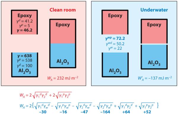
    1. Clint JH, Wicks AC. Adhesion under water: surface energy considerations. Int. J. Adhes. Adhes. 2001;21:267–73.
    1. Pocius AV. Adhesives and Adhesives Technology. Hanser/Gardner Publ.; Cincinnati: 1997.

LinkOut - more resources