. 2011 Oct 31:2:77.
doi: 10.3389/fphys.2011.00077. eCollection 2011.

Engineering Parvalbumin for the Heart: Optimizing the Mg Binding Properties of Rat β-Parvalbumin

Affiliations

Engineering Parvalbumin for the Heart: Optimizing the Mg Binding Properties of Rat β-Parvalbumin

Jianchao Zhang et al. Front Physiol. .

Abstract

Parvalbumin (PV), an EF-hand protein family member, is a delayed calcium buffer that exchanges magnesium for calcium to facilitate fast skeletal muscle relaxation. Genetic approaches that express parvalbumin in the heart also enhance relaxation and show promise of being therapeutic against various cardiac diseases where relaxation is compromised. Unfortunately, skeletal muscle PVs have very slow rates of Ca(2+) dissociation and are prone to becoming saturated with Ca(2+), eventually losing their buffering capability within the constantly beating heart. In order for PV to have a more therapeutic potential in the heart, a PV with faster rates of calcium dissociation and high Mg(2+) affinity is needed. We demonstrate that at 35°C, rat β-PV has an ~30-fold faster rate of Ca(2+) dissociation compared to rat skeletal muscle α-PV, and still possesses a physiologically relevant Ca(2+) affinity (~100 nM). However, rat β-PV will not be a delayed Ca(2+) buffer since its Mg(2+) affinity is too low (~1 mM). We have engineered two mutations into rat β-PV, S55D and E62D, when observed alone increase Mg(2+) affinity up to fivefold, but when combined increase Mg(2+) affinity ~13-fold, well within a physiologically relevant affinity. Furthermore, the Mg(2+) dissociation rate (172/s) from the engineered S55D, E62D PV is slow enough for delayed Ca(2+) buffering. Additionally, the engineered PV retains a high Ca(2+) affinity (132 nM) and fast rate of Ca(2+) dissociation (64/s). These PV design strategies hold promise for the development of new therapies to remediate relaxation abnormalities in different heart diseases and heart failure.

Keywords: calcium; magnesium; parvalbumin; relaxation.


Figures

Figure 1
Figure 1
Purification of PV. (A) SDS-PAGE analysis of the DEAE column purification profile for the E62D, F102W β-PV. The gel was stained with Coomassie Brilliant Blue. Lane 1: a positive control sample from rat F102W β-PV, which was characterized before the nucleic acid contamination issue was resolved. Lanes 2–15 correspond to elution fractions from 21 to 35 after the DEAE column (refer to Materials and Method). (B) the same SDS-PAGE from (A), but was re-stained with Ethidium Bromide and viewed with UV light (refer to Materials and Methods).
Figure 2
Figure 2
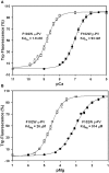
Ca2+ and Mg2+ binding to the F102W α-PV and F102W β-PV. (A) The Ca2+ dependent increase in Trp fluorescence is shown as a function of −Log[Ca2+] (pCa) for F102W α-PV (□) and F102W β-PV (•). Increasing concentrations of Ca2+ were added to 1 μM protein in 2 ml of 200 mM MOPS, 150 mM KCl, 4 mM EGTA, pH 7.0 at 35°C. Trp fluorescence was monitored at 330 nm with excitation at 295 nm. Each data point represents the mean ± SE of at least three titrations. (B) The Mg2+ dependent increase in Trp fluorescence is shown as a function of −Log[Mg2+] (pMg) for F102W α-PV (□) and F102W β-PV (•). The experimental conditions were the same as described for (A).
Figure 3
Figure 3
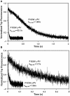
Rates of Ca2+ and Mg2+ dissociation from F102W α-P and F102W β-PV. (A) The time course of Trp fluorescence is shown as EDTA rapidly chelates Ca2+ causing dissociation of Ca2+ from F102W α-PV and F102W β-PV. Each protein (5 μM) in 10 mM MOPS, 150 mM KCl, 10 μM Ca2+, pH 7.0 at 35°C was rapidly mixed with equal volume of 30 mM EDTA in 10 mM MOPS, 150 mM KCl, pH 7.0. (B) The time course of Trp fluorescence is shown as EDTA rapidly chelates Mg2+ causing dissociation of Mg2+ from F102W α-PV and F102W β-PV. Each protein (5 μM) in 10 mM MOPS, 150 mM KCl, 5 mM EGTA, 500 μM Mg2+, pH 7.0 at 35°C was rapidly mixed with equal volume of 30 mM EDTA in 10 mM MOPS, 150 mM KCl, pH 7.0. Trp fluorescence was monitored through a narrow band-pass filter centered at 334 nm with an excitation wavelength of 295 nm. Each trace is an average of at least five traces fit with a single exponential equation. All kinetic traces were triggered at time zero.
Figure 4
Figure 4
Ca2+ and Mg2+ binding and dissociation from S55D, F102W β-PV. (A) The Ca2+ dependent increase in Trp fluorescence is shown as a function of −Log[Ca2+] (pCa) for S55D, F102W β-PV. (B) The time course of Trp fluorescence is shown as EDTA rapidly chelates Ca2+ causing dissociation of Ca2+ from S55D, F102W β-PV. (C) The Mg2+ dependent increase in Trp fluorescence is shown as a function of −Log[Mg2+] (pMg) for S55D, F102W β-PV. (D) The time course of Trp fluorescence is shown as EDTA rapidly chelates Mg2+ causing dissociation of Mg2+ from S55D, F102W β-PV. All the measurements were performed as previously mentioned in the legends of Figures 2 and 3.
Figure 5
Figure 5
Ca2+ and Mg2+ binding and dissociation from E62D, F102W β-PV. (A) The Ca2+ dependent increase in Trp fluorescence is shown as a function of −Log[Ca2+] (pCa) for E62D, F102W β-PV. (B) The time course of Trp fluorescence is shown as EDTA rapidly chelates Ca2+ causing dissociation of Ca2+ from E62D, F102W β-PV. (C) The Mg2+ dependent increase in Trp fluorescence is shown as a function of −Log[Mg2+] (pMg) for E62D, F102W β-PV. (D) The time course of Trp fluorescence is shown as EDTA rapidly chelates Mg2+ causing dissociation of Mg2+ from E62D, F102W β-PV. All the measurements were performed as previously mentioned in the legends of Figures 2 and 3.
Figure 6
Figure 6
Ca2+ and Mg2+ binding and dissociation from S55D, E62D, F102W β-PV. (A) The Ca2+ dependent increase in Trp fluorescence is shown as a function of −Log[Ca2+] (pCa) for S55D, E62D, F102W β-PV. (B) The time course of Trp fluorescence is shown as EDTA rapidly chelates Ca2+ causing dissociation of Ca2+ from S55D, E62D, F102W β-PV. (C) The Mg2+ dependent increase in Trp fluorescence is shown as a function of −Log[Mg2+] (pMg) for S55D, E62D, F102W β-PV. (D) The time course of Trp fluorescence is shown as EDTA rapidly chelates Mg2+ causing dissociation of Mg2+ from S55D, E62D, F102W β-PV. All the measurements were performed as previously mentioned in the legends of Figures 2 and 3.

References

    1. Arif S. H. (2009). A Ca(2+)-binding protein with numerous roles and uses: parvalbumin in molecular biology and physiology. Bioessays 31, 410–42110.1002/bies.200800170 - DOI - PubMed
    1. Belkacemi L., Simoneau L., Lafond J. (2002). Calcium-binding proteins: distribution and implication in mammalian placenta. Endocrine 19, 57–6410.1385/ENDO:19:1:57 - DOI - PubMed
    1. Bers D. M. (2006). Altered cardiac myocyte Ca regulation in heart failure. Physiology (Bethesda) 21, 380–38710.1152/physiol.00019.2006 - DOI - PubMed
    1. Cates M. S., Berry M. B., Ho E. L., Li Q., Potter J. D., Phillips G. N., Jr. (1999). Metal-ion affinity and specificity in EF-hand proteins: coordination geometry and domain plasticity in parvalbumin. Structure 7, 1269–127810.1016/S0969-2126(00)80060-X - DOI - PubMed
    1. Csillik B., Schwaller B., Mihaly A., Henzi T., Losonczi E., Knyihar-Csillik E. (2010). Upregulated expression of oncomodulin, the beta isoform of parvalbumin, in perikarya and axons in the diencephalon of parvalbumin knockout mice. Neuroscience 165, 749–75710.1016/j.neuroscience.2009.10.048 - DOI - PubMed