

Phase-shift, stimuli-responsive drug carriers for targeted delivery
- PMID: 22059114
- PMCID: PMC3207214
- DOI: 10.4155/tde.11.81
Phase-shift, stimuli-responsive drug carriers for targeted delivery
Abstract
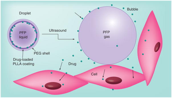
The intersection of particles and directed energy is a rich source of novel and useful technology that is only recently being realized for medicine. One of the most promising applications is directed drug delivery. This review focuses on phase-shift nanoparticles (that is, particles of submicron size) as well as micron-scale particles whose action depends on an external-energy triggered, first-order phase shift from a liquid to gas state of either the particle itself or of the surrounding medium. These particles have tremendous potential for actively disrupting their environment for altering transport properties and unloading drugs. This review covers in detail ultrasound and laser-activated phase-shift nano- and micro-particles and their use in drug delivery. Phase-shift based drug-delivery mechanisms and competing technologies are discussed.
Figures


Similar articles
-
Phase-shift, stimuli-responsive perfluorocarbon nanodroplets for drug delivery to cancer.Wiley Interdiscip Rev Nanomed Nanobiotechnol. 2012 Sep-Oct;4(5):492-510. doi: 10.1002/wnan.1176. Epub 2012 Jun 22. Wiley Interdiscip Rev Nanomed Nanobiotechnol. 2012. PMID: 22730185 Free PMC article. Review.
-
Drug-Loaded Perfluorocarbon Nanodroplets for Ultrasound-Mediated Drug Delivery.Adv Exp Med Biol. 2016;880:221-41. doi: 10.1007/978-3-319-22536-4_13. Adv Exp Med Biol. 2016. PMID: 26486341 Review.
-
Design and physicochemical characterization of advanced spray-dried tacrolimus multifunctional particles for inhalation.Drug Des Devel Ther. 2013;7:59-72. doi: 10.2147/DDDT.S40166. Epub 2013 Feb 4. Drug Des Devel Ther. 2013. PMID: 23403805 Free PMC article.
-
Margination of micro- and nano-particles in blood flow and its effect on drug delivery.Sci Rep. 2014 May 2;4:4871. doi: 10.1038/srep04871. Sci Rep. 2014. PMID: 24786000 Free PMC article.
-
Redox-responsive nano-carriers as tumor-targeted drug delivery systems.Eur J Med Chem. 2018 Sep 5;157:705-715. doi: 10.1016/j.ejmech.2018.08.034. Epub 2018 Aug 13. Eur J Med Chem. 2018. PMID: 30138802 Review.
Cited by
-
Microfluidic generation of acoustically active nanodroplets.Small. 2012 Jun 25;8(12):1876-9. doi: 10.1002/smll.201102418. Epub 2012 Mar 29. Small. 2012. PMID: 22467628 Free PMC article.
-
Current status and prospects for microbubbles in ultrasound theranostics.Wiley Interdiscip Rev Nanomed Nanobiotechnol. 2013 Jul-Aug;5(4):329-45. doi: 10.1002/wnan.1219. Epub 2013 Mar 15. Wiley Interdiscip Rev Nanomed Nanobiotechnol. 2013. PMID: 23504911 Free PMC article. Review.
-
On-demand intracellular amplification of chemoradiation with cancer-specific plasmonic nanobubbles.Nat Med. 2014 Jul;20(7):778-784. doi: 10.1038/nm.3484. Epub 2014 Jun 1. Nat Med. 2014. PMID: 24880615 Free PMC article.
-
Ultrasound-triggered phase transition sensitive magnetic fluorescent nanodroplets as a multimodal imaging contrast agent in rat and mouse model.PLoS One. 2013 Dec 31;8(12):e85003. doi: 10.1371/journal.pone.0085003. eCollection 2013. PLoS One. 2013. PMID: 24391983 Free PMC article.
-
Current advances in ultrasound-combined nanobubbles for cancer-targeted therapy: a review of the current status and future perspectives.RSC Adv. 2021 Apr 6;11(21):12915-12928. doi: 10.1039/d0ra08727k. eCollection 2021 Mar 29. RSC Adv. 2021. PMID: 35423829 Free PMC article. Review.
References
Bibliography
-
- Hahn MA, Singh AK, Sharma P, Brown SC, Moudgil BM. Nanoparticles as contrast agents for in-vivo bioimaging: current status and future perspectives. Anal Bioanal Chem. 2011;399(1):3–27. - PubMed
-
- Pan D, Lanza GM, Wickline SA, Caruthers SD. Nanomedicine: perspective and promises with ligand-directed molecular imaging. Eur J Radiol. 2009;70(2):274–285. - PubMed
Patent
-
- Apfel RE. Activatable infusible dispersions containing drops of a superheated liquid for methods of therapy and diagnosis. 5840276. US Patent. 1998
Publication types
MeSH terms
Substances
Grants and funding
LinkOut - more resources
Full Text Sources