. 2011 Sep;23(6):669-694.
doi: 10.1080/20445911.2011.594433. Epub 2011 Sep 22.

Neural correlates of emotion-cognition interactions: A review of evidence from brain imaging investigations

Affiliations
Free PMC article

Neural correlates of emotion-cognition interactions: A review of evidence from brain imaging investigations

Florin Dolcos et al. J Cogn Psychol (Hove). 2011 Sep.
Free PMC article

Abstract

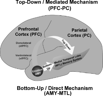
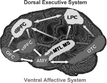
Complex dynamic behaviour involves reciprocal influences between emotion and cognition. On the one hand, emotion is a "double-edged sword" that may affect various aspects of our cognition and behaviour, by enhancing or hindering them and exerting both transient and long-term influences. On the other hand, emotion processing is also susceptible to cognitive influences, typically exerted in the form of emotion regulation. Noteworthy, both of these reciprocal influences are subjective to individual differences that may affect the way we perceive, experience, and eventually remember emotional experiences, or respond to emotionally challenging situations. Understanding these relationships is critical, as unbalanced emotion-cognition interactions may lead to devastating effects, such as those observed in mood and anxiety disorders. The present review analyses the reciprocal relationships between emotion and cognition, based on evidence derived from brain imaging investigations focusing on three main topics: (1) the impact of emotion on cognition, (2) the impact of cognition on emotion, and (3) the role of individual differences in emotion-cognition interactions. This evidence will be discussed in the context of identifying aspects that are fundamental to understanding the mechanisms underlying emotion-cognition interactions in healthy functioning, and to understanding changes associated with affective disorders.


Figures

Figure 1.
Figure 1.
Main mechanisms involved in the memory-enhancing effect of emotion. Emotion enhances long-term episodic memory by modulating activity in two main memory-related brain regions, the medial–temporal lobe (MTL) memory system and the prefrontal cortex (PFC). However, the effects of emotion on MTL and PFC regions may be related to different mechanisms: AMY and MTL are part of basic/direct neurohormonal mechanisms underlying the memory-enhancement effect of emotion (bottom-up mechanism), whereas PFC is part of a mechanism (also including the parietal cortex—PC) that has an indirect/mediated involvement in the formation of emotional memories, by enhancing strategic, semantic, working memory, and attentional processes (top–down mechanism). Adapted from LaBar and Cabeza (2006); courtesy of Dr. Roberto Cabeza. [To view this figure in colour, please visit the online version of this Journal.]
Figure 2.
Figure 2.
Neural systems involved in cognitive/executive (dorsal) versus emotional (ventral) processing. The dorsal system includes brain regions typically associated with “cold” executive (ColdEx) colour–coded in blue) functions, such as the dorsolateral prefrontal cortex (dlPFC) and the lateral parietal cortex (LPC), which are critical to active maintenance of goal-relevant information in working memory (WM). The ventral system includes brain regions involved in “hot” emotional (HotEmo; colour–coded in red) processing, such as the amygdala (AMY), the ventrolateral PFC (vlPFC), and the medial PFC. Other brain regions that these systems interact with (MTL MS, OTC) are also illustrated. MTL MS = medial temporal lobe memory system, OFC = orbitofrontal cortex, OTC = occipitotemporal cortex. Monochromatic arrows represent connections within the same system, whereas bichromatic arrows represent connections across systems. Although visual cortical areas illustrated here (OTC) are not technically part of the HotEmo system, they are coloured in red because these areas are susceptible to influences from emotion processing regions. This representation does not include all regions that are part of the two systems, as in its present format it does not include medial brain regions. Adapted from figure provided by Dr. Lihong Wang. [To view this figure in colour, please visit the online version of this Journal.]
Figure 3.
Figure 3.
Evidence for the role of lateral PFC in coping with distracting emotions. (A) Brain regions showing enhanced functional coupling with the amygdala during processing of emotional distraction—ventrolateral prefrontal cortex (vlPFC)/inferior frontal cortex (IFC) highlighted. (B) Enhanced correlation between vlPFC activity and subjective emotional distractibility scores. (C) Hemispheric asymmetry in the vlPFC/IFC during successful coping with emotional distraction. Taken together, these findings can also be interpreted as a subjective (right) versus objective (left) dissociation in the role of these regions in coping with the subjective feeling of being distracted (right vlPFC/IFC) versus actually coping with distraction (left vlPFC/IFC). Correct/Incorrect Remembered/Forgotten items in the working memory task. Adapted from Dolcos, Kragel, et al. (2006) and Dolcos and McCarthy (2006), with permission. [To view this figure in colour, please visit the online version of this Journal.]
Figure 4.
Figure 4.
Sex-related differences identified in the amygdala showing a hemispheric asymmetry in memory-related activity. (A) Activity in the left amygdala while viewing emotionally arousing images was more significantly related to subsequent memory for the images in women than in men, and (B) the opposite pattern was observed in the right amygdala, i.e., greater activity in men than in women. Reproduced from Cahill et al. (2004), with permission. [To view this figure in colour, please visit the online version of this Journal.]
Figure 5.
Figure 5.
Preserved amygdala function and increased amygdala–ACC connectivity in healthy ageing. (A) Coronal brain view and bar graph showing overlapping activity in the right AMY region during processing of negative pictures (Neg > Neu) in young and older adults. The y–axis represents the difference in activity between negative and neutral conditions and units are in effect size, the difference in the parameter estimates of the activation. (B) Sagittal/lateral brain view and bar graph showing age-related increase in the interaction between AMY and the anterior cingulate cortex (ACC) during processing of negative pictures. The y–axis represents the difference in correlations between negative and neutral conditions. From St. Jacques et al. (2010), with permission. [To view this figure in colour, please visit the online version of this Journal.]

References

    1. Adolphs R. The social brain: Neural basis of social knowledge. Annual Review of Psychology. 2009;60:693–716. - PMC - PubMed
    1. Allen P. A., Kaut K. P., Baena E., Lien M.-C., Ruthruff E. Individual differences in positive affect moderate age-related declines in episodic long-term memory. Journal of Cognitive Psychology. 2011;23(6):768–779.
    1. Allen P. A., Kaut K. P., Lord R. G., Hall R. J., Grabbe J. W., Bowie T. An emotional mediation theory of differential age effects in episodic and semantic memories. Experimental Aging Research. 2005;31(4):355–391. - PubMed
    1. Amaral D. G., Price J. L., Pitkanen A., Carmichael S. T. The amygdala: Neurobiological aspects of emotion, memory, and mental dysfunction. New York, NY: Wiley-Liss; 1992.
    1. Anderson A. K., Phelps E. A. Lesions of the human amygdala impair enhanced perception of emotionally salient events. Nature. 2001;411:305–309. - PubMed

LinkOut - more resources