

Autofocus methods of whole slide imaging systems and the introduction of a second-generation independent dual sensor scanning method
- PMID: 22059145
- PMCID: PMC3205523
- DOI: 10.4103/2153-3539.86282
Autofocus methods of whole slide imaging systems and the introduction of a second-generation independent dual sensor scanning method
Abstract
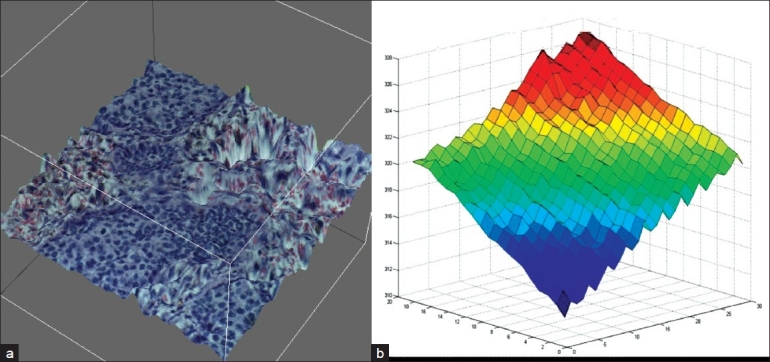
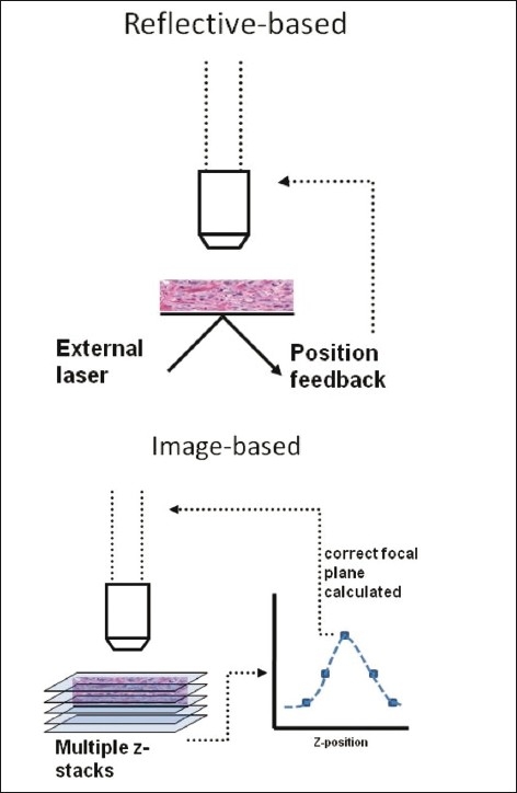
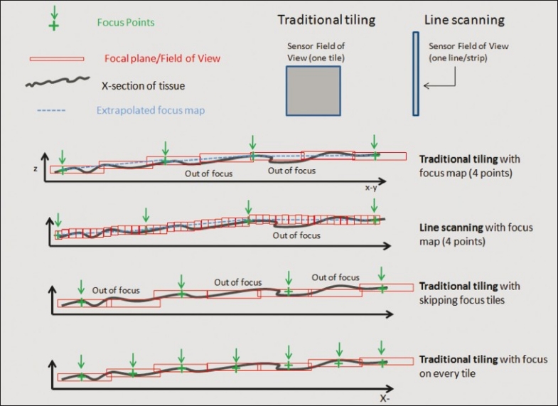
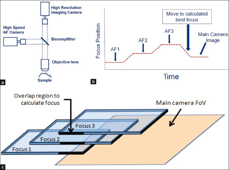
Accurate focusing is a critical challenge of whole slide imaging, primarily due to inherent tissue topography variability. Traditional line scanning and tile-based scanning systems are limited in their ability to acquire a high degree of focus points while still maintaining high throughput. This review examines limitations with first-generation whole slide scanning systems and explores a novel approach that employs continuous autofocus, referred to as independent dual sensor scanning. This "second-generation" concept decouples image acquisition from focusing, allowing for rapid scanning while maintaining continuous accurate focus. The technical concepts, merits, and limitations of this method are explained and compared to that of a traditional whole slide scanning system.
Keywords: Autofocus; digital pathology; whole slide imaging.
Figures
Similar articles
-
Autofocusing technologies for whole slide imaging and automated microscopy.J Biophotonics. 2020 Dec;13(12):e202000227. doi: 10.1002/jbio.202000227. Epub 2020 Sep 18. J Biophotonics. 2020. PMID: 32844560 Review.
-
The accuracy of dynamic predictive autofocusing for whole slide imaging.J Pathol Inform. 2011;2:38. doi: 10.4103/2153-3539.84231. Epub 2011 Aug 24. J Pathol Inform. 2011. PMID: 21969919 Free PMC article.
-
High throughput slanted scanning whole slide imaging system for digital pathology.J Biophotonics. 2021 Jun;14(6):e202000499. doi: 10.1002/jbio.202000499. Epub 2021 Mar 8. J Biophotonics. 2021. PMID: 33638313
-
Dynamic autofocus for continuous-scanning time-delay-and-integration image acquisition in automated microscopy.J Biomed Opt. 2007 May-Jun;12(3):034011. doi: 10.1117/1.2743078. J Biomed Opt. 2007. PMID: 17614719
-
Applications and challenges of digital pathology and whole slide imaging.Biotech Histochem. 2015 Jul;90(5):341-7. doi: 10.3109/10520295.2015.1044566. Epub 2015 May 15. Biotech Histochem. 2015. PMID: 25978139 Review.
Cited by
-
Rapid and robust whole slide imaging based on LED-array illumination and color-multiplexed single-shot autofocusing.Quant Imaging Med Surg. 2019 May;9(5):823-831. doi: 10.21037/qims.2019.05.04. Quant Imaging Med Surg. 2019. PMID: 31281778 Free PMC article.
-
Whole-Slide Image Focus Quality: Automatic Assessment and Impact on AI Cancer Detection.J Pathol Inform. 2019 Dec 12;10:39. doi: 10.4103/jpi.jpi_11_19. eCollection 2019. J Pathol Inform. 2019. PMID: 31921487 Free PMC article.
-
Removing defocused objects from single focal plane scans of cytological slides.J Pathol Inform. 2016 May 4;7:21. doi: 10.4103/2153-3539.181765. eCollection 2016. J Pathol Inform. 2016. PMID: 27217971 Free PMC article.
-
Terapixel hyperspectral whole-slide imaging via slit-array detection and projection.J Biomed Opt. 2018 Jun;23(6):1-7. doi: 10.1117/1.JBO.23.6.066503. J Biomed Opt. 2018. PMID: 29959834 Free PMC article.
-
Single-frame rapid autofocusing for brightfield and fluorescence whole slide imaging.Biomed Opt Express. 2016 Oct 27;7(11):4763-4768. doi: 10.1364/BOE.7.004763. eCollection 2016 Nov 1. Biomed Opt Express. 2016. PMID: 27896014 Free PMC article.
References
-
- Firestone L, Cook K, Culp K, Talsania N, Preston K., Jr Comparison of autofocus methods for automated microscopy. Cytometry. 2004;12:195–206. - PubMed
-
- Sun Y, Duthaler S, Nelson BJ. Autofocusing in computer microscopy: Selecting the optimal focus algorithm. Micros Res Tech. 2004;65:139–49. - PubMed
-
- Rojo MG, García GB, Mateos CP, García JG, Vicente MC. Critical comparison of 31 commercially available digital slide systems in pathology. Int J Surg Pathol. 2006;14:285–305. - PubMed
-
- Yazdanfar S, Kenny KB, Tasimi K, Corwin AD, Dixon EL, Filkins RJ. Simple and robust image-based autofocusing for digital microscopy. Opt Express. 2008;16:8670–7. - PubMed
LinkOut - more resources
Full Text Sources
Other Literature Sources
