

High-definition hematoxylin and eosin staining in a transition to digital pathology
- PMID: 22059146
- PMCID: PMC3205517
- DOI: 10.4103/2153-3539.86284
High-definition hematoxylin and eosin staining in a transition to digital pathology
Abstract
Introduction: A lot of attention has been generated in recent years by digital pathology and telepathology. Multiple reasons for and barriers to effective adoption are discussed in the current literature. Digital slides are the most promising medium at this time. The goal of our study was to evaluate whether the change in the methodology, particularly utilizing the so-called high-definition hematoxylin and eosin (H and E) slides, enhanced the quality of the final digital slide, and whether pathologists who tested the results perceived this as a difference in quality.
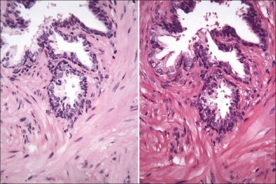
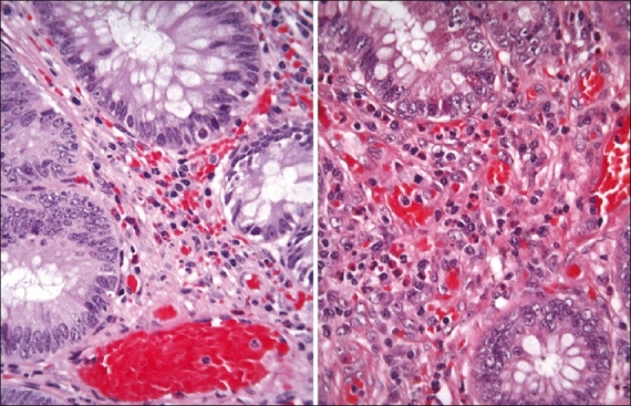
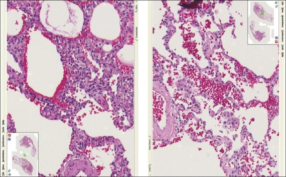
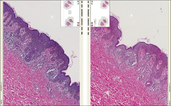
Methods: THE STUDY WAS A BLINDED COMPARISON OF DIGITAL SLIDES PREPARED USING TWO METHODS: standard H&E batch staining and automated individual "high definition" HD HE staining. Four pathologists have compared 80 cases stained with each method.
Results: The results discussed in this study show potential promise that the utilization of protocol(s) adapted for tissue and for imaging might be preferable for digital pathology in at least some of the pathology subspecialties. In particular, the protocol evaluated here was capable of turning out digital slides that had more contrast and detail, and therefore were perceived to provide enhanced diagnostically significant information for the pathologist.
Keywords: Digital pathology; digital slides; histochemistry; histology; imaging; staining methods; staining protocols; telepathology.
Figures
References
-
- Jara-Lazaro AR, Thamboo TP, Teh M, Tan PH. Digital pathology: exploring its applications in diagnostic surgical pathology practice. Pathology. 2010;42:512–8. - PubMed
-
- Al-Janabi S, Huisman A, Van Diest PJ. Digital pathology: current status and future perspectives. Histopathology. 2011 doi: 10.1111/j.1365-2559.2011.03814.x. - PubMed
-
- Jukić DM, Drogowski LM, Martina J, Parwani AV. Clinical examination and validation of primary diagnosis in anatomic pathology using whole slide digital images. Arch Pathol Lab Med. 2011;135:372–8. - PubMed
LinkOut - more resources
Full Text Sources
Research Materials
