

Evaluation and optimization for liquid-based preparation cytology in whole slide imaging
- PMID: 22059147
- PMCID: PMC3205520
- DOI: 10.4103/2153-3539.86285
Evaluation and optimization for liquid-based preparation cytology in whole slide imaging
Abstract
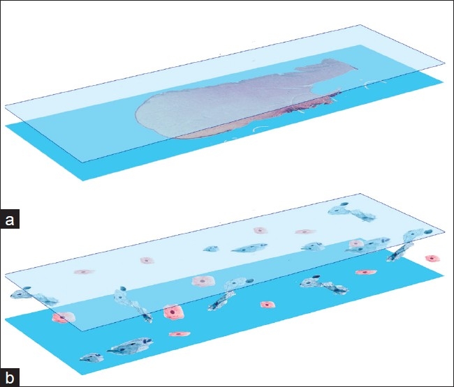
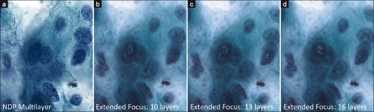
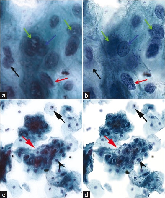
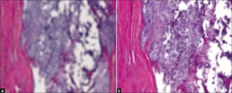
Background: Cytology poses different obstacles in whole slide imaging compared to surgical pathology slides. A single focal plane suffices for most of the latter, but cytology slides are thicker, potentially requiring multiple focal planes for adequate diagnostic information. Multiple focal planes adversely impact scanning time per slide, evaluation times, and file sizes. In this pilot study, we evaluated and compared the multilayer stack method to the extended focus algorithm as an alternative which collapses multiple focal planes into a single image, retaining only focused areas from each plane.
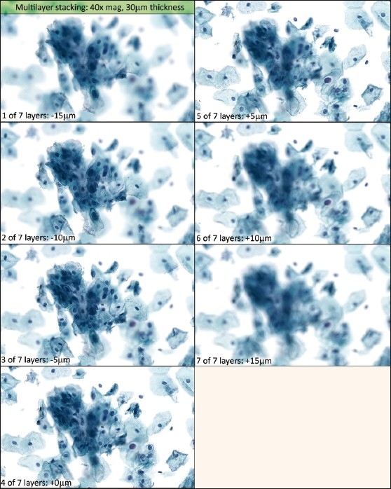
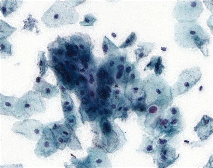
Materials and methods: 10 SurePath(®) cervical cytology slides were scanned at three thickness settings: 18, 24, and 30 μm. Three scanners were used: (1) Hamamatsu Nanozoomer 2.0-HT, (2) 3DHISTECH Mirax scan, and (3) Bioimagene iScan Coreo Au. The Nanozoomer and iScan utilized multilayer stacking, while the Mirax files were composited by extended focus. Scan times and file sizes were recorded, and image quality compared.
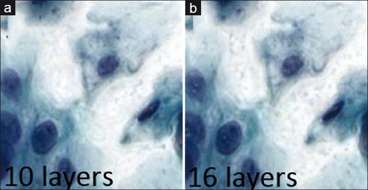
Results: The Nanozoomer stacks averaged 1.58 gb and around 25 min for each slide, while the iScan stacks ranged from 6.23 to 9.3 gb and took 34-50 min to scan. The Mirax images averaged 210 mb and took 13-20 min to scan. Multilayer stack image quality from both Nanozoomer and iScan was fairly comparable. The iScan revealed significant mechanical issues that did not correspond to user settings. The Mirax images showed worrisome loss of crisp focus detail, worsening with increasing focal planes and impacting assessment of nuclear contours and chromatin detail.
Conclusions: The optimal number of focal planes remains unknown for cytology. Multilayer stacks require excessive scanning time, network bandwidth, and file storage. Extended focus was evaluated as an alternative, but significant image quality issues were revealed. Further large-scale studies are needed to assess their clinical impact.
Keywords: Cytology; cytopathology; digital; pathology; whole slide imaging.
Figures
References
-
- Evered A, Dudding N. Accuracy and perceptions of virtual microscopy compared with glass slide microscopy in cervical cytology. Cytopathology. 2010 May 17; DOI: 101111/j1365-2303201000758x. - PubMed
-
- Solomon D, Nayar R. The Bethesda System for Reporting Cervical Cytology. 2nd ed. New York: Springer Science + Business Media LLC; 2001. p. 4.
-
- Wilbur DC. Digital cytology: current State of the art and prospects for the future. Acta Cytol. 2011;55:227–38. - PubMed
-
- Qayyum S, Yagi Y, Wilbur DC. Optimization of Whole Slide Imaging Parameters for Liquid-Based Cervical Cytology Slides. Presented at 57th Annual Scientific Meeting of the American Society of Cytopathology in 2009
LinkOut - more resources
Full Text Sources
Miscellaneous
