

Usefulness of endoscopic ultrasonography in hepatology
- PMID: 22059170
- PMCID: PMC3222772
- DOI: 10.1155/2011/367643
Usefulness of endoscopic ultrasonography in hepatology
Abstract
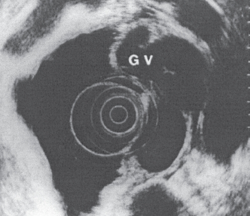
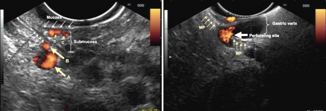
Endoscopic ultrasonography (EUS) is used to evaluate patients with hepatobiliary diseases. The technique is useful for the diagnosis of esogastric varices in selected cases of portal hypertension, and to evaluate the pathogenic role and prognostic value of the collateral circulation in patients with this condition. When coupled with the Doppler technique, EUS can be used to guide injection sclerotherapy and to verify the obliteration of varices (particularly fundal varices) after endoscopic treatment. Hemodynamic changes induced in the collateral circulation by vasoactive drugs can also be measured with Doppler-EUS. Fine-needle aspiration under EUS guidance is useful in the diagnosis of focal liver lesions and perihepatic adenopathy, and in the evaluation of biliary tract diseases. New indications can be developed in the future after adequate experimental validation.
L’échographie per endoscopique (EE) est un nouvel outil utilisé pour évaluer les patients ayant une suspicion de maladie hépato-biliaire. Elle est utile pour le diagnostic des varices oeso-gastriques dans certains cas particuliers d’hypertension portale; elle permet aussi d’étudier la circulation collatérale, sa signification physiopathologique et sa valeur pronostique. L’association de l’EE avec la technique Doppler permet de guider la sclérothérapie de varices per endoscopique et également de vérifier l’oblitération des varices après traitement (particulièrement les varices fundiques). Il est également possible de mesurer les effets hémodynamiques de médicaments vaso-actifs sur la circulation collatérale. La cytologie à l’aiguille fine peut être réalisée grâce à un guidage échoendoscopique au niveau des lésions hépatiques focales ou d’adénopathies suspectes ainsi que de lésions de l’arbre biliaire. Il est probable que de nouvelles applications vont émerger après une validation expérimentale adéquate.
Figures

References
-
- Ginès A, Fernandez-Esprrach G. Endoscopic ultrasonography for the evaluation of portal hypertension. Clin Liver Dis. 2010;14:221–9. - PubMed
-
- De Lisi S, Buscarini E. Endoscopic ultrasonography and portal hypertension: Where are we in 2009 ? Eur J Gastroenterol Hepatol. 2009;21:1327–32. - PubMed
-
- Sgouros SN, Vasiliadis KV, Pereira SP. Systematic review: Endoscopic and imaging-based techniques in the assessment of portal haemodynamics and the risk of variceal bleeding. Aliment Pharmacol Ther. 2009;30:965–76. - PubMed
-
- Baik SK. Hemodynamic evaluation by Doppler ultrasonography in patients with portal hypertension: A review. Liver Int. 2010;30:1403–13. - PubMed
-
- Lee YT, Chan FKL, Ching JYL, et al. Diagnosis of gastroesophageal varices and portal collateral venous abnormalities by endosonography in cirrhotic patients. Endoscopy. 2002;34:391–8. - PubMed
Publication types
MeSH terms
LinkOut - more resources
Full Text Sources
Medical