. 2011 Dec;82(5):1150-63.
doi: 10.1111/j.1365-2958.2011.07875.x. Epub 2011 Nov 7.

Sulfoquinovose synthase - an important enzyme in the N-glycosylation pathway of Sulfolobus acidocaldarius

Affiliations

Sulfoquinovose synthase - an important enzyme in the N-glycosylation pathway of Sulfolobus acidocaldarius

Benjamin H Meyer et al. Mol Microbiol. 2011 Dec.

Abstract

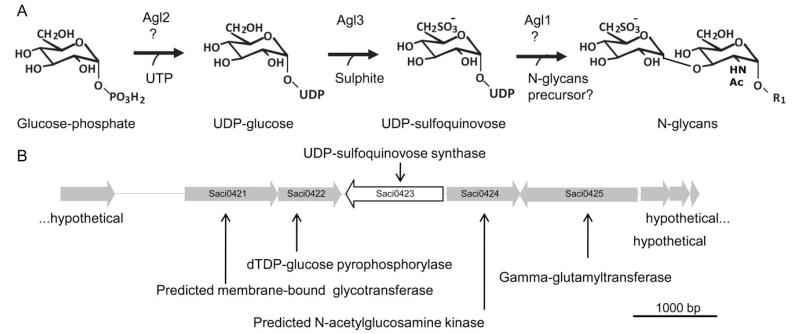
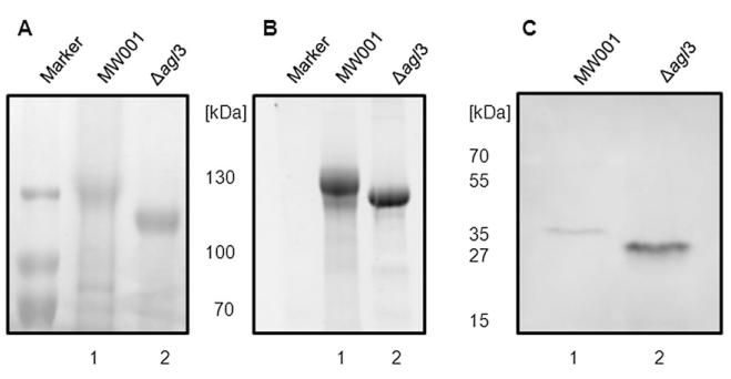
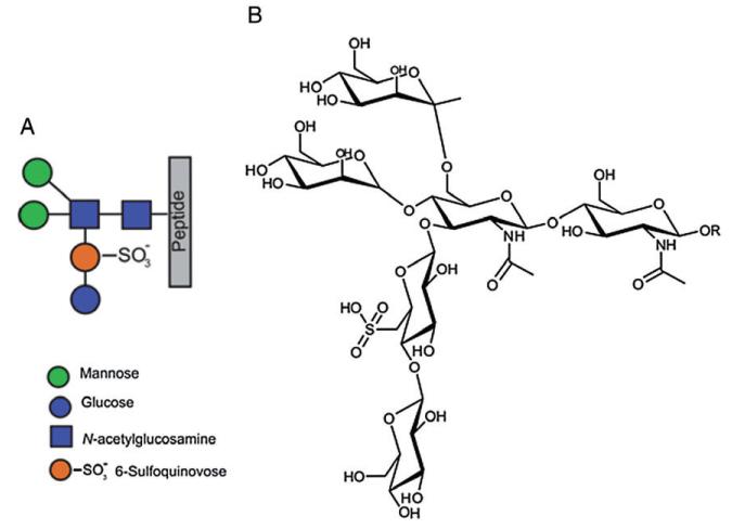
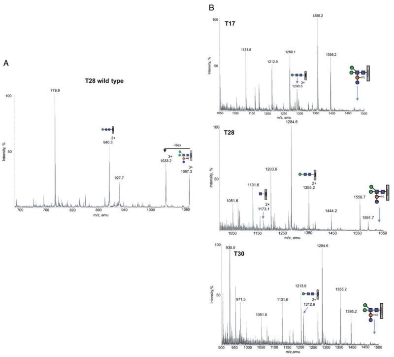
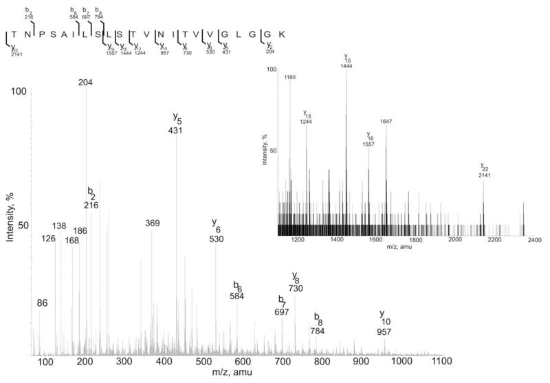
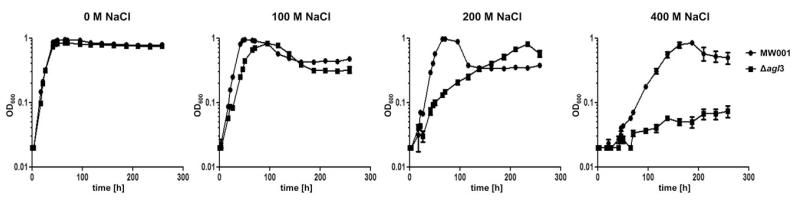
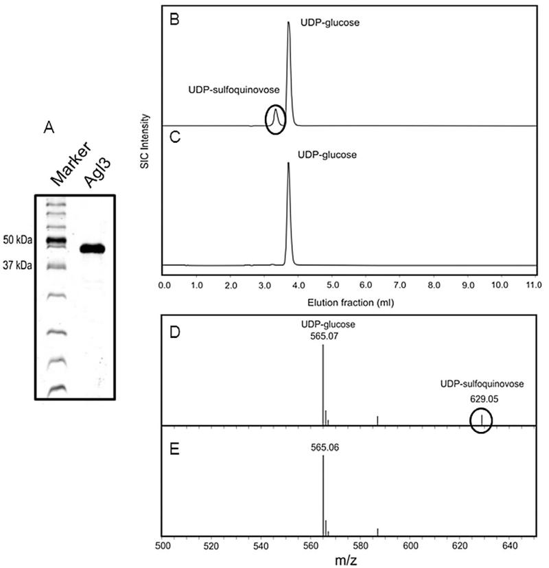
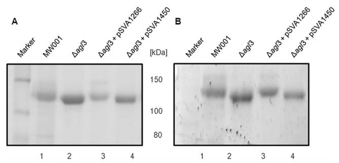
Recently, the Surface (S)-layer glycoprotein of the thermoacidophilic crenarchaeote Sulfolobus acidocaldarius was found to be N-glycosylated with a heterogeneous family of glycans, with the largest having a composition Glc(1)Man(2)GlcNAc(2) plus 6-sulfoquinovose. However, genetic analyses of genes involved in the N-glycosylation process in Crenarchaeota were missing so far. In this study we identify a gene cluster involved in the biosynthesis of sulfoquinovose and important for the assembly of the S-layer N-glycans. A successful markerless in-frame deletion of agl3 resulted in a decreased molecular mass of the S-layer glycoprotein SlaA and the flagellin FlaB, indicating a change in the N-glycan composition. Analyses with nanoLC ES-MS/MS confirmed the presence of only a reduced trisaccharide structure composed of Man(1) GlcNAc(2) , missing the sulfoquinovose, a mannose and glucose. Biochemical studies of the recombinant Agl3 confirmed the proposed function as a UDP-sulfoquinovose synthase. Furthermore, S. acidocaldarius cells lacking agl3 had a significantly lower growth rate at elevated salt concentrations compared with the background strain, underlining the importance of the N-glycosylation to maintain an intact and stable cell envelope, to enable the survival of S. acidocaldarius in its extreme environment.


Figures

Fig. 1
Fig. 1
Probable sulfoquinovose biosynthesis pathway in S. acidocaldarius. A. Proposed biosynthetic pathway of the UDP-sulfoquinovose in S. acidocaldarius. R1, GlcNAc-P-Dol; UTP, uridine triphosphate; agl2 encoding for an UDP-glucose pyrophosphorylase; agl3 coding for an UDP-sulfoquinovose synthase; agl1 coding for a membrane bound glucosyltransferase. Biosynthetic pathway in agreement with (Shimojima, 2011). B. Physical map of the gene region of S. acidocaldarius, in which the gene coding for UDP-sulfoquinovose synthase is located. Illustrated are the genes Saci0420 until Saci0428. The red displayed gene encodes the UDP-sulfoquinovose synthase. The genes Saci0421 und Saci0422 are most likely also involved in the biosynthesis of sulfoquinovose (see A).
Fig. 2
Fig. 2
Confirmation of the in-frame deletion mutant Δagl3 (Saci0423). A. Gene deletion was confirmed by PCR using the outside primers against the flanking regions of agl3, DNA isolated from the background strain MW001 (lane 2), plasmid pSVA1225 used for the homologous recombination incorporating the up- and downstream region of deleted agl3 (lane 3), or the mutant lacking agl3 (lane 1). B. RT-PCR confirms the deletion of the gene agl3 presumably encoding the UDP-sulfoquinovose. The cDNA (C) served as a template in PCR amplification using primers against the internal region of either the agl3 or aglB. In each case, PCR amplifications were also performed using genomic DNA (G) as a positive control and total RNA (R) as a negative control.
Fig. 3
Fig. 3
Effect of the agl3 deletion on S. acidocaldarius S-layer protein glycosylation. Equivalent amounts of the S-layer protein SlaA isolated from S. acidocaldarius MW001 (lane 1) and Δagl3 (lane 2) were separated by 8% SDS-PAGE and either (A) Coomassie blue-stained or (B) stained for carbohydrates using Emerald 300 (B). C. Effect on S. acidocaldarius flagella protein FlaB glycosylation of the agl3 deletion. Equivalent amounts of cells from S. acidocaldarius MW001 (lane 1) and Δagl3 (lane 2) were separated by 11% SDS-PAGE and immunoblotted with antibodies raised against FlaB.
Fig. 4
Fig. 4
Graphic and chemical representation of the S-layer N-glycan. A. The full glycan structure shown by the colour-coded symbols. B. Chemical structure of the Glc1Man2GlcNAc2QuiS hexasaccharide observed in the wild type S. acidocaldarius.
Fig. 5
Fig. 5
Detailed analysis of the Δagl3 deletion mutant S-layer glycop peptide. A. Total ion chromatogram of glycan profile for the T28 peptide in S. acidocaldarius wild type with the annotated peaks showing the glycoforms observed (M+3H)3+. B. Total ion chromatogram for three selected glycopeptides in S. acidocaldarius lacking agl3. The annotated peaks show the Hex1HexNAc2 glycoform (M+2H)2+ is preserved on all glycopeptides, whereas the full glycan is completely missing. For reference, each spectra is annotated with the absent full glycan for each glycopeptide.
Fig. 6
Fig. 6
MSMS data of the T28 glycopeptide with the Hex1HeNAc2 residue. Both b and y theoretical ions firmly matched with the experimental MSMS data. Inset figure shows MS spectra with larger m/z-values.
Fig. 7
Fig. 7
Response to salt stress in the Δagl3 strain. S. acidocaldarius MW001 or Δagl3 were grown in Brock medium at different salt concentrations (0–400 mM NaCl). Growth was measured at an optical density of 600 nm (OD600). Shown are values obtained from two independent repeats.
Fig. 8
Fig. 8
Agl3 is a UDP-sulfoquinovose synthase. A. SDS-PAGE analysis (12% gel) of the expression and purification of sulfoquinovose synthase (Agl3) from S. acidocaldarius in E. coli. B and C. RP-HPLC analysis of the conversion of UDP-glucose and sulphite into UDP-sulfoquinovose catalysed by (B) Agl3 and the (C) negative control. D and E. PGC-ESI-MS analysis showing the conversion of UDP-glucose and sulphite into UDP-sulfoquinovose catalysed by Agl3 (D) and negative control (E).
Fig. 9
Fig. 9
Complementation of the Δagl3 deletion mutant. Equivalent amounts of the S-layer protein SlaA isolated from S. acidocaldarius MW001 (lane 1), Δagl3 (lane 2), Δagl3 complemented with pSVA1266 (lane 3), and Δagl3 with control plasmid pSVA1450 (lane 4) were separated by 8% SDS-PAGE and either (A) Coomassie blue-stained or (B) stained for carbohydrates using Emerald 300 (B).

References

    1. Abu-Qarn M, Yurist-Doutsch S, Giordano A, Trauner A, Morris HR, Hitchen P, et al. Haloferax volcanii AglB and AglD are involved in N-glycosylation of the S-layer glycoprotein and proper assembly of the surface layer. J Mol Biol. 2007;374:1224–1236. - PubMed
    1. Abu-Qarn M, Giordano A, Battaglia F, Trauner A, Hitchen PG, Morris HR, et al. Identification of AglE, a second glycosyltransferase involved in N- glycosylation of the Haloferax volcanii S-layer glycoprotein. J Bacteriol. 2008;190:3140–3146. - PMC - PubMed
    1. Albers SV, Meyer BH. The archaeal cell envelope. Nat Rev Microbiol. 2011;9:414–426. - PubMed
    1. Albers SV, Elferink MG, Charlebois RL, Sensen CW, Driessen AJ, Konings WN. Glucose transport in the extremely thermoacidophilic Sulfolobus solfataricus involves a high-affinity membrane-integrated binding protein. J Bacteriol. 1999;181:4285–4291. - PMC - PubMed
    1. Benning C. Biosynthesis and function of the sulfolipid sulfoquinovosyl diacylglycerol. Annu Rev Plant Physiol Plant Mol Biol. 1998;49:53–75. - PubMed

Publication types

MeSH terms