Review
. 2012 Jan 17;84(2):779-92.
doi: 10.1021/ac202812h. Epub 2011 Nov 7.

Interferometric methods for label-free molecular interaction studies

Affiliations
Review

Interferometric methods for label-free molecular interaction studies

Amanda Kussrow et al. Anal Chem. .
No abstract available


Figures

Figure 1
Figure 1
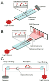
Block diagrams of (A) a Mach–Zehnder interferometer (MZI), (B) a Young interferometer (YI), and (C) a Hartman interferometer (HI).
Figure 2
Figure 2
(A) Overlay of normalized analyte binding zones for human IgG captured by immobilized Protein A. Reprinted from ref , Copyright 2010, with permission from Elsevier. (B) Overlay of representative sample of normalized analyte binding zones for quantitation of a 46 kDa fusion protein. Reprinted from ref , Copyright 2010, with permission from Elsevier. (C) Schematic diagram of the three geometries that can be used to obtain diffraction images. Reprinted from ref . Copyright 2009 American Chemical Society.
Figure 3
Figure 3
(A) Schematic of the dual-polarization interferometer. Reprinted from ref , Copyright 2004, with permission from Elsevier. (B) The optical extinction during the injection of 10 μM methylene blue over the DNA/PEI layer (a), the DNA/amino layer (b), bare chip surface (d), the PEI surface (e), and the amino surface (f), or 10 μM mitoxantrone over the DNA/amino layer (c) and the amino surface (g). The RI and mass of the DNA layer on PEI: 1.382, 2.05 ng/mm2; the RI and mass of the DNA layer on amino surface: 1.379, 1.36 ng/mm2. Reprinted from ref . Copyright 2010 American Chemical Society.
Figure 4
Figure 4
(A) Waveguide microresonator geometries. The resonance path is highlighted in red; blue indicates microfluidic flow. Reprinted by permission from Macmillian Publishers Ltd: Nature Methods, ref , Copyright 2008. (B) Real time response curves for 5 representative sensors (out of 20 total) and corresponding calibration curves based on the initial slope of the response curve. For the response curves, the colored lines represent the following concentrations of the antibody-specific antigen: red, 150 ng/mL; green, 100 ng/mL; blue, 50 ng/mL; purple, 20 ng/mL; orange, 10 ng/mL; black, 0 ng/mL; gray, unknown solutions. For the calibration curves, black circles are the calibration points and red squares represent the unknowns. Reprinted from ref . Copyright 2010 American Chemical Society.
Figure 5
Figure 5
Block diagram of a porous silicon sensor.
Figure 6
Figure 6
(A) Block diagram of backscattering interferometry (BSI). (B) Cartoon illustration of the fringe shift seen upon binding and the resulting BSI signal.
Figure 7
Figure 7
(A) BSI shows the difference in binding affinities for free-solution and surface-immobilized DNA hybridization. (B) BSI detects the kinetic interaction of calmodulin and trifluoperazine. From ref . Reprinted with permission from American Association for the Advancement of Science, Copyright 2007. (C) Shows that the magnitude of binding as detected by BSI for α-crystallin to different T4L lysozyme mutants. Reprinted from ref . Copyright 2009 American Chemical Society. (D) Quantitative measurement of titer strength for syphilis in human sera samples. Ref –Reproduced by permission of The Royal Society of Chemistry, Copyright 2010. (E) Binding of trinitrophenol to its antibody using an end point assay. Reprinted with permission from ref . Copyright 2011 John Wiley & Sons, Inc. (F) Binding of GABA to its receptor in a native cell membrane. Reprinted with permission from ref . Copyright 2011 Nature Publishing Group.

References

    1. Herschel W. Philos Trans R Soc, London. 1805;95:31–64.
    1. Baldwin JE, Haniff CA. Philos Trans: Math, Phys Eng Sci. 2002;360:969–986. - PubMed
    1. Michelson AA, Pease FG. Astrophys J. 1921;53:249–259.
    1. Bruning JH, Herriott DR, Gallaghe Je, Rosenfel Dp, White AD, Brangacc DJ. Appl Opt. 1974;13:2693–2703. - PubMed
    1. Scudieri F. Appl Opt. 1980;19:404–408. - PubMed

Publication types