. 2012 Jan 6;415(1):175-92.
doi: 10.1016/j.jmb.2011.10.003. Epub 2011 Oct 31.

Computational design of high-affinity epitope scaffolds by backbone grafting of a linear epitope

Affiliations

Computational design of high-affinity epitope scaffolds by backbone grafting of a linear epitope

Mihai L Azoitei et al. J Mol Biol. .

Abstract

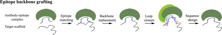
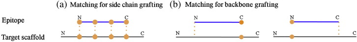
Computational grafting of functional motifs onto scaffold proteins is a promising way to engineer novel proteins with pre-specified functionalities. Typically, protein grafting involves the transplantation of protein side chains from a functional motif onto structurally homologous regions of scaffold proteins. Using this approach, we previously transplanted the human immunodeficiency virus 2F5 and 4E10 epitopes onto heterologous proteins to design novel "epitope-scaffold" antigens. However, side-chain grafting is limited by the availability of scaffolds with compatible backbone for a given epitope structure and offers no route to modify backbone structure to improve mimicry or binding affinity. To address this, we report here a new and more aggressive computational method-backbone grafting of linear motifs-that transplants the backbone and side chains of linear functional motifs onto scaffold proteins. To test this method, we first used side-chain grafting to design new 2F5 epitope scaffolds with improved biophysical characteristics. We then independently transplanted the 2F5 epitope onto three of the same parent scaffolds using the newly developed backbone grafting procedure. Crystal structures of side-chain and backbone grafting designs showed close agreement with both the computational models and the desired epitope structure. In two cases, backbone grafting scaffolds bound antibody 2F5 with 30- and 9-fold higher affinity than corresponding side-chain grafting designs. These results demonstrate that flexible backbone methods for epitope grafting can significantly improve binding affinities over those achieved by fixed backbone methods alone. Backbone grafting of linear motifs is a general method to transplant functional motifs when backbone remodeling of the target scaffold is necessary.


Figures

None
Graphical abstract
Fig. 1
Fig. 1
Main stages of epitope side-chain and backbone grafting protocols. Given a starting antibody–epitope complex, candidate scaffolds (red) are identified in the matching stage for epitope (yellow) transplantation. In epitope side-chain grafting, the residue identity of the candidate scaffold is altered to match the epitope sequence. In epitope backbone grafting, the epitope backbone conformation replaces the native backbone region of the candidate scaffold; the  2F5 mAb is shown in blue (heavy chain) and magenta (light chain).
Fig. 2
Fig. 2
Models of representative epitope scaffolds designed by side-chain grafting and chosen for experimental characterization. The scaffolds are shown in red with the epitope in yellow; the  2F5 mAb is in blue (heavy chain) and magenta (light chain).
Fig. 3
Fig. 3
SPR analysis of the  2F5 mAb interactions of selected epitope scaffolds and epitope peptides. (a–c) Binding responses of epitope peptides of different lengths. (d and g) 2F5 binding of corresponding epitope scaffolds designed by side-chain and backbone grafting (e and h). (f and i) Equilibrium binding analysis and affinity comparison of epitope peptides and epitope scaffolds designed by the two different computational protocols; the collected data are shown in black and the model fits are shown in red (1:1 Langmuir binding) and blue (conformational change model).
Fig. 4
Fig. 4
Thermal stability of corresponding epitope scaffolds designed by side-chain and backbone grafting.
Fig. 5
Fig. 5
Stages of epitope backbone grafting. (I) The epitope is aligned on the target scaffold. (II) The native scaffold backbone corresponding to the epitope is deleted, resulting in a disconnected polypeptide chain. (III) To integrate the epitope with the scaffold, novel backbone regions are modeled between the epitope termini and the scaffold (red stars). (IV) Final closure of the chain sets the rigid-body orientation of the epitope and the antibody relative to the scaffold; sequence design ensures the stabilization of the epitope conformation and the productive interaction of the antibody with the resulting epitope scaffold.
Fig. 6
Fig. 6
Comparison of epitope-scaffold models generated by side-chain and backbone grafting based on the same native protein (PDB ID: 1wnu). (a) Alignment of side-chain grafting (red) and the backbone grafting models (blue) in complex with 2F5 (shown in gray when interacting with the backbone grafting design and in blue and magenta in complex with the side-chain grafting design). Changes in the orientation of the epitope region (yellow) relative to the scaffold alter the binding orientation of 2F5. (b) Detailed view (black box) of the different epitope conformations. The central DKW epitope residues are shown in stick representation. (c) Changes in the conformation of the scaffold backbone in the epitope region allow the addition of extra epitope residues (P50L) as well as the engineering of stabilizing interaction between the epitope and the scaffold (I58M) in the backbone grafting model.
Fig. 7
Fig. 7
Crystal structure analysis of epitope scaffolds and comparison with the computational models. Epitope regions are shown in detailed view. (a) Alignment of the SC_2cx5 crystal structure (yellow) and the computational model (red). (b) Alignment of the BB_2cx5_001 crystal structure (blue) and the computational model (red). (c) Alignment of the SC_2cx5 (yellow) and BB_2cx5_001 (blue) structures. (d) Mutated residues between SC_2cx5 (yellow) and BB_2cx5_001 mapped on the respective structures; the mutated residues and the DKW epitope motif are shown as sticks; SC_2cx5 residues are shown in salmon and BB_2cx5_001 residues are shown in cyan. (e) Alignment of the SC_1wnu crystal structure (green) and the computational model (red). (f) Alignment of the BB_1wnu_001 crystal structure (magenta) and the computational model (red). (g) Alignment of the SC_1wnu (green) and BB_1wnu_001 (magenta) structures. (h) Mutated residues between SC_1wnu (green) and BB_1wnu_001 (magenta) mapped on the respective structures; the mutated residues and the DKW epitope motif are shown as sticks; SC_1wnu residues are shown in orange and BB_2cx5_001 residues are shown in blue.
Fig. S1
Fig. S1
Surface plasmon resonance analysis of epitope scaffolds and epitope peptides binding to 2F5 mAb. The collected data are shown in black and the model fits are shown in red (1:1 Langmuir binding) and blue (conformational change model). The concentrations of the injected samples are shown in the kinetic analysis panels.
Fig. S2
Fig. S2
Wavelength scans and thermal stability of epitope scaffolds.
Fig. S3
Fig. S3
Sequence alignment of epitope scaffolds designed by epitope side chain grafting (SC_2cx5 and SC_1wnu) and epitope backbone grafting (BB_2cx5_001–003 and BB_1wnu_001–003); the epitope sequence is shown in red; sequences changes in blue were introduced to transplant the epitope onto the parent scaffolds by side-chain grafting; sequence changes in green indicated differences between the models generated by epitope side-chain and epitope backbone grafting; additional epitope residues introduced during epitope backbone grafting are shown in magenta.
Fig. S4
Fig. S4
Epitope-mediated crystal contacts and B-factors in the determined crystal structures of epitope scaffolds; the epitope scaffolds are shown in red, with the epitope in yellow; the epitope is shown in stick representation except for the tryptophan residue (equivalent to W666 of gp41), which is shown in sphere representation; symmetry-related molecules are shown in gray.
Fig. S5
Fig. S5
Matching schemes for epitope matching. (a) The full epitope structure is aligned over the target scaffold in epitope side-chain grafting. (b) Only one of the epitope ends is aligned on target scaffolds in epitope backbone grafting; orange circles indicate pairs of residues on the epitope and scaffold for which backbone RMSD is evaluated.

References

    1. Kritzer J.A., Zutshi R., Cheah M., Ran F.A., Webman R., Wongjirad T.M., Schepartz A. Miniature protein inhibitors of the p53–hDM2 interaction. ChemBioChem. 2006;7:29–31. - PubMed
    1. Cobos E.S., Pisabarro M.T., Vega M.C., Lacroix E., Serrano L., Ruiz-Sanz J., Martinez J.C. A miniprotein scaffold used to assemble the polyproline II binding epitope recognized by SH3 domains. J. Mol. Biol. 2004;342:355–365. - PubMed
    1. Sia S.K., Kim P.S. Protein grafting of an HIV-1-inhibiting epitope. Proc. Natl Acad. Sci. USA. 2003;100:9756–9761. - PMC - PubMed
    1. Chin J.W., Schepartz A. Concerted evolution of structure and function in a miniature protein. J. Am. Chem. Soc. 2001;123:2929–2930. - PMC - PubMed
    1. Zanghellini A., Jiang L., Wollacott A.M., Cheng G., Meiler J., Althoff E.A. New algorithms and an in silico benchmark for computational enzyme design. Protein Sci. 2006;15:2785–2794. - PMC - PubMed

Publication types

MeSH terms

LinkOut - more resources