. 2012 Feb;45(2):733-42.
doi: 10.1016/j.nbd.2011.10.020. Epub 2011 Oct 29.

Inhibition of prolyl hydroxylases by dimethyloxaloylglycine after stroke reduces ischemic brain injury and requires hypoxia inducible factor-1α

Affiliations

Inhibition of prolyl hydroxylases by dimethyloxaloylglycine after stroke reduces ischemic brain injury and requires hypoxia inducible factor-1α

Molly E Ogle et al. Neurobiol Dis. 2012 Feb.

Abstract

Pathological oxygen deprivation inhibits prolyl hydroxylase (PHD) activity and stimulates a protective cellular oxygen-sensing response in part through the stabilization and activation of the Hypoxia Inducible Factor (HIF) 1α transcription factor. The present investigation tested the therapeutic potential of enhanced activation of oxygen-sensing pathways by competitive pharmacologic PHD inhibition after stroke, hypothesizing that post-ischemic PHD inhibition would reduce neuronal cell death and require the activation of HIF-1α. The PHD inhibitor dimethyloxaloylglycine (DMOG, 100 μM) reduced cell death by oxygen glucose deprivation (OGD), an in vitro model of ischemia, and the protection required HIF-1α. In vivo, DMOG (50 mg/kg, i.p.) administered 30 or 60 min after distal occlusion of the middle cerebral artery (MCA) in mice enhanced the activation of HIF-1α protein, enhanced transcription of the HIF-regulated genes vascular endothelial growth factor, erythropoietin, endothelial nitric oxide synthase, and pyruvate dehydrogenase kinase-1, reduced ischemic infarct volume and activation of the pro-apoptotic caspase-3 protein, reduced behavioral deficits after stroke, and reduced the loss of local blood flow in the MCA territory after stroke. Inhibition of HIF-1α in vivo by Digoxin or Acriflavine abrogated the infarct sparing properties of DMOG. These data suggest that supplemental activation of oxygen-sensing pathways after stroke may provide a clinically applicable intervention for the promotion of neurovascular cell survival after ischemia.


Conflict of interest statement

Conflict of interest: N/A

Figures

Fig 1
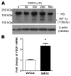
Fig 1. DMOG induces normoxic HIF-1α expression in cortical neurons
A. Primary cortical neurons were treated with indicated concentration of DMOG for 24h. Western blot demonstrates an increase in HIF-1α (115 kDa) protein expression with DMOG treatment. β-actin is a loading control. B. Neurons were treated for 24h with vehicle or 250μM DMOG. Quantitative RT-PCR was used to determine the expression of HIF-1-dependent gene VEGF. (n=3, Mean ± SEM, * p<0.05).
Fig 2
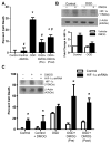
Fig 2. PHD inhibitor attenuates OGD-induced cell death and requires HIF-1α
Cortical neurons were subjected to OGD for 2h with 24h reperfusion. Cell death was assessed by trypan blue exclusion 24h after OGD. Cortical neurons were pre-treated 24h prior to OGD or post-treated after OGD with 100μM DMOG. (A) DMOG reduced cell death in both pre- and post- treatment. (*p<0.05 compared to control; #p<0.05 compared to OGD; n=4). (B) Western blot of protein collected from cortical neurons 3h after OGD or OGD + DMOG demonstrate that HIF-1α is enhanced after OGD in the DMOG treatment group (n=3). (C) Inset demonstrates successful HIF-1α knockdown. Neuronal cultures were infected with lentiviral shRNA directed against HIF-1α for 48h. Parallel control and HIF-1α-shRNA cells were treated with 250μM DMOG for 24h to induce HIF-1α protein stabilization. Control cultures but not shRNA treated cultures show HIF-1α protein expression. DMOG did not reduce cell death from OGD in cells with HIF-1α knockdown. Data are the mean ± SEM from at least three independent experiments. (*compared to parallel control cultures, p<0.05, n=3).
Fig 3
Fig 3. PHD inhibitor DMOG attenuates apoptotic cell death in cortical neurons
(A) Pure neuronal cultures were subjected to an apoptotic insult of B-27 supplement withdrawal for 24h and dead cells were counted by trypan blue staining. (B) Neuronal lysate was collected after 24h and immunoblotted for HIF-1α and β-actin loading control. (C) Western blot for the caspase-3 active fragment 3 and 6h after initiation of B-27 withdrawal. Data are the mean ± SEM from at least three independent experiments. (*p<0.05; n=3).
Fig 4
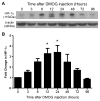
Fig 4. DMOG intraperitoneal injection stabilizes HIF-1α protein in the brain
(A) Adult male mice were injected with 50mg/kg DMOG and sacrificed at the indicated time intervals (hours). Cortical tissue was harvested and immunoblotted for HIF-1α or β actin. Representative western is shown. (B) Densitometry summary of western blots. (n=3–4 animals per time-point; mean ± SEM, *compared to vehicle injected animal, p<0.05).
Fig 5
Fig 5. PHD inhibitor post-ischemic treatment reduces ischemic infarct formation and attenuates peri-infarct loss of cerebral perfusion
Representative TTC staining of ischemic infarct 72h after stroke in (A) control and (B) 60 min DMOG (50mg/kg) post-treatment. (C) Quantification of TTC infarct volume (mm3) 72h after stroke demonstrates the protective effect of PHD inhibitor post-ischemic treatment. (n=10–12 animals per group, *p<0.05). (D) Local cerebral blood flow (CBF) was examined over the territory of the right middle cerebral artery covering the ischemic border region in our focal ischemic stroke model. (E) Pseudo-colored representation of intensity of CBF before, during, and 72h after stroke in control or DMOG post-treated animals. (F) Quantification of CBF relative to initial flow. (n=8, *p<0.05).
Fig 6
Fig 6. PHD inhibitor treatment reduced activation of caspase-3 in the ischemic cortex
(A) Immuno-staining for active caspase-3 and neuronal nuclei (NeuN) 24h after ischemia. (B) Quantification of the number of active caspase-3 positive cells in the peri-infarct region. DMOG post-treatment reduced the number of apoptotic neurons. (n=4, *p<0.05).
Fig 7
Fig 7. PHD inhibitor post-ischemic treatment reduces sensorimotor behavioral deficits after stroke
Adhesive removal test for sensorimotor forelimb function before, 1–3, 7 and 14 days after stroke. Deficits are measured as increased amount of time to remove the adhesive dot in the affected left forepaw (A) and the unaffected right forepaw (B). (*p<0.05, n=10–12 animals per group)
Fig 8
Fig 8. Post-ischemic DMOG therapy enhances HIF-1α expression
(A) Expression of HIF-1α 3h after stroke with vehicle or DMOG 30 min post-treatment. (* p<0.05, n=4) (B) mRNA expression of HIF-1 responsive genes in control stroke animals or DMOG-treated animals 12h after stroke. (* p<0.05, n=3–4)
Fig 9
Fig 9. Inhibition of HIF-1α abrogates the post-ischemic DMOG-mediated neuroprotection
(A) Adult male mice were pre-treated for 6h with saline or HIF-1α inhibitors 2mg/kg DIG, or 2mg/kg ACF. Mice in each group were then treated with DMOG (50mg/kg) or vehicle. After 24h, cortical tissues were immunoblotted for HIF-1α. Densitometry is represented as fold change to vehicle for each condition. DMOG enhances HIF-1α expression compared to vehicle in control conditions, but not under DIG treatment. ACF shows a trend for enhanced expression of HIF-1α with DMOG, but it is not statistically significant. (n=3 animals per group). (B) RNA was extracted from cortical tissues treated with DIG, ACF, and DMOG. Quantitative RT-PCR for the HIF-1-dependent gene VEGF illustrates that DIG and ACF inhibit the DMOG-induced transcription of HIF-1 dependent VEGF expression. (C) Mice were pre-treated for 24h with saline, DIG, or ACF and then underwent surgical dMCAO. Mice in each group were treated 30m after stroke with either DMOG or vehicle. After 12h, peri-infarct cortical tissue was immunoblotted for HIF-1α and actin loading control. DMOG enhances HIF-1α expression compared to sham in control conditions; DIG and ACF reduce the DMOG-mediated increase in HIF-1a. Quantification of densitometry is fold change compared to sham control. (n=3–4, p<0.05) (D) Quantitative RT-PCR analysis of VEGF and EPO from peri-infarct cortical tissue 12h after stroke shows that DIG and ACF reduce the DMOG-mediated increase in expression. All data are fold change relative to sham control. (n=3–4 animals per group, p<0.05) (E) Mice were pre-treated for 24h with HIF inhibitors DIG or ACF. TTC staining of ischemic infarct 72h after stroke with or without DMOG post-treatment shows no protection in the HIF-1α inhibitor groups. (F) Quantification of infarct volume. (n=8–10 in each group, * p<0.05).

References

    1. Abu-Amara M, Yang SY, Quaglia A, Rowley P, Fuller B, Seifalian A, Davidson B. Role of endothelial nitric oxide synthase in remote ischemic preconditioning of the mouse liver. Liver Transpl. 2011;17:610–619. - PubMed
    1. American Heart Association. Heart Disease and Stroke Statistics. Dallas, Texas, USA: American Heart Association; (2008 Update)
    1. Aminova LR, Chavez JC, Lee J, Ryu H, Kung A, Lamanna JC, Ratan RR. Prosurvival and prodeath effects of hypoxia-inducible factor-1alpha stabilization in a murine hippocampal cell line. J Biol Chem. 2005;280:3996–4003. - PubMed
    1. Aragones J, et al. Deficiency or inhibition of oxygen sensor Phd1 induces hypoxia tolerance by reprogramming basal metabolism. Nat Genet. 2008;40:170–180. - PubMed
    1. Baranova O, Miranda LF, Pichiule P, Dragatsis I, Johnson RS, Chavez JC. Neuron-specific inactivation of the hypoxia inducible factor 1 alpha increases brain injury in a mouse model of transient focal cerebral ischemia. J Neurosci. 2007;27:6320–6332. - PMC - PubMed

Publication types

MeSH terms