. 2012 Feb;1821(2):268-78.
doi: 10.1016/j.bbalip.2011.10.017. Epub 2011 Oct 29.

Distinct cellular pools of perilipin 5 point to roles in lipid trafficking

Affiliations

Distinct cellular pools of perilipin 5 point to roles in lipid trafficking

Sadie R Bartholomew et al. Biochim Biophys Acta. 2012 Feb.

Abstract

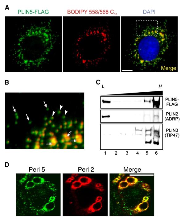
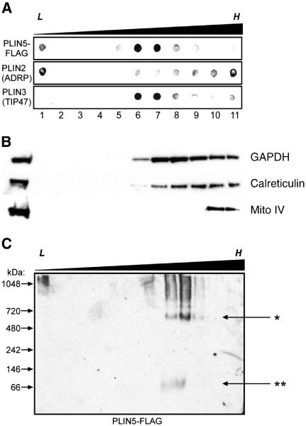
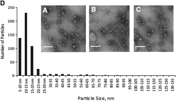
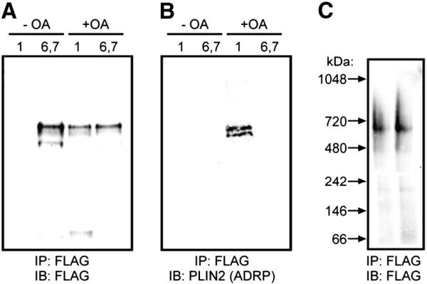
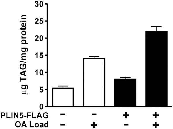
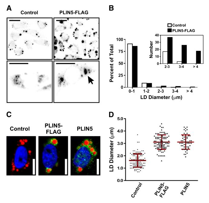
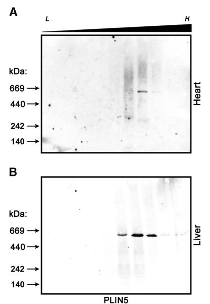
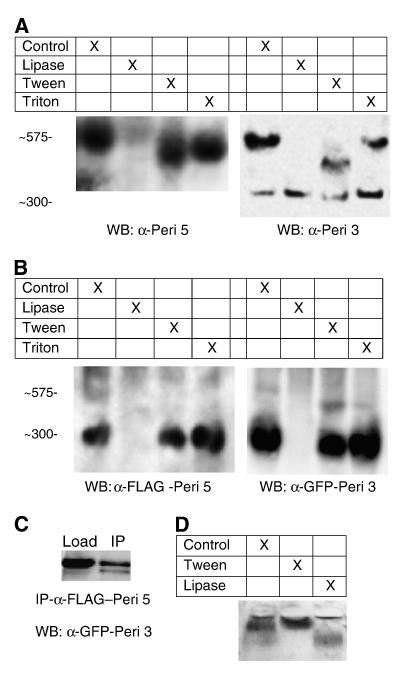
The PAT family of lipid storage droplet proteins comprised five members, each of which has become an established regulator of cellular neutral lipid metabolism. Perilipin 5 (also known as lsdp-5, MLDP, PAT-1, and OXPAT), the most recently discovered member of the family, has been shown to localize to two distinct intracellular pools: the lipid storage droplet (LD), and a poorly characterized cytosolic fraction. We have characterized the denser of these intracellular pools and find that a population of perilipin 5 not associated with large LDs resides in complexes with a discrete density (~1.15 g/ml) and size (~575 kDa). Using immunofluorescence, western blotting of isolated sucrose density fractions, native gradient gel electrophoresis, and co-immunoprecipitation, we have shown that these small (~15 nm), perilipin 5-encoated structures do not contain the PAT protein perilipin 2 (ADRP), but do contain perilipin 3 and several other as of yet uncharacterized proteins. The size and density of these particles as well as their susceptibility to degradation by lipases suggest that like larger LDs, they have a neutral lipid rich core. When treated with oleic acid to promote neutral lipid deposition, cells ectopically expressing perilipin 5 experienced a reorganization of LDs in the cell, resulting in fewer, larger droplets at the expense of smaller ones. Collectively, these data demonstrate that a portion of cytosolic perilipin 5 resides in high density lipid droplet complexes that participate in cellular neutral lipid accumulation.


Figures

Fig. 1
Fig. 1
Perilipin 5 localizes to biochemically distinct intracellular pools. A. Confocal images of a representative CHO cell stably expressing the perilipin 5-3X-FLAG construct (green). The perilipin 5-3X-FLAG protein was detected using an anti-FLAG antibody (green). Neutral lipids were stained through incorporation of BODIPY 558/568 C12 (red), and nuclei were stained with DAPI (blue). Scale bar = 10 μm. B. Inset from merged image (right panel in A). Under basal growth conditions, perilipin 5-3X-FLAG localized to both LDs (double arrows) as well as smaller punctate structures in the cytosol. Of the latter, some were associated with neutral lipid staining (arrowheads), while others were apparently devoid of BODIPY 558/568 C12 labeling (arrows). C. Perilipin 5-3X-FLAG-expressing CHO cell lysates prepared following incubation in lipid-loading medium, were subjected to ultracentrifugation using conventional discontinuous sucrose density gradients (0–30%). Immunoblots were prepared using aliquots of equal volume fractions of the gradients, arranged from low (L) to high (H) density, and probed with anti-FLAG, anti-perilipin 2 (ADRP), and anti-perilipin 3 (TIP47) antibodies. Whereas the majority of perilipin 2 and perilipin 3 partitioned into either low or high-density fractions, respectively, perilipin 5-3X-FLAG was recovered in fractions at both extremes of the density gradient. D. Confocal images of a representative CHO cell stably expressing the perilipin 5-3X-FLAG construct (green) and stained with an anti-perilipin 2 antibody (red). Merged images are shown in yellow. Both perilipin 5 and perilipin 2 localize to lipid storage droplets but only perilipin 5 is found on punctate structures.
Fig. 2
Fig. 2
A portion of perilipin 5 is associated with high-density structures of a discrete size. A. Perilipin 5-FLAG-expressing CHO cell lysates were subjected to ultracentrifugation using extended discontinuous sucrose density gradients (0–60%). Dot blots were prepared using aliquots of equal volume fractions of the gradients, arranged from low (L) to high (H) density, and probed with anti-FLAG, anti-perilipin 2 (ADRP), and anti-perilipin 3 (TIP47) antibodies. When the gradients were extended over a wider density range, perilipin 5 was found to partition into both low-density (buoyant) fractions (lane 1) as well as fractions of higher density (lanes 6–7), but was essentially absent in the fractions of highest density (lanes 9–11). B. Marker proteins for cytosol (NADPH), ER (calreticulin) and mitochondria (mito complex IV) are shown. C. Non-denaturing gradient gel electrophoresis was performed using sucrose gradient fractions prepared as described in A. Immunoblots were probed with anti-FLAG antibodies. Immunoreactive material that partitioned into the denser fractions of the gradient also migrated into the gel under non-denaturing conditions, revealing an approximate size of 575 kDa (marked by the asterisk, *). A small amount of immunoreactivity was found migrating near the predicted size of perilipin 5 (double asterisk, **). Please note that each panel contains representative data from different experiments.
Fig. 3
Fig. 3
A majority of high-density perilipin 5-containing structures have a size of less than 20 nm. Transmission electron microscopy was performed on material immunoprecipitated from the post-nuclear supernatant isolated from perilipin 5-3X-FLAG-expressing CHO cells grown under basal conditions. A–C Representative images of negative stained immunoprecipitated fractions. Scale bars = 100 nm. D. Histogram showing the size distribution of 500 particles from randomly chosen, non-overlapping microscopic fields, as determined using morphometric analysis.
Fig. 4
Fig. 4
Co-immunoprecipitation of perilipin 5 coated particles confirms a lack of perilipin 2/ADRP in high-density structures. In A–C, cells were either treated with oleic acid (+ OA) or basal growth conditions (− OA). Sucrose gradient (0–60%) ultracentrifugation was performed and fractions 1 (low density) or 6 and 7 (high density) were immunoprecipitated with anti-FLAG beads. The eluted material was subjected to SDS-PAGE and either prepared for immunoblot analysis (A, B), or developed using silver staining, as noted. D. Immunoprecipitated particles from CHO cells were eluted with FLAG peptide and analyzed by non-denaturing gradient gel electrophoresis followed by immunoblotting and detection using anti-FLAG antibodies. No shift in the size of these ~ 575 kDa particles was observed following immunoprecipitation.
Fig. 5
Fig. 5
CHO fibroblasts expressing perilipin 5 store more TAG than control cells. CHO cells expressing the perilipin-5 3-X-FLAG fusion protein or transfected with a control plasmid were assayed for cellular TAG in the presence or absence of exogenous oleic acid (OA) treatment for 24 h (n = 6). TAG data were normalized to total protein concentration (mean ± SEM). Relative to control cells, perilipin 5 expressors stored 0.7-fold more TAG under basal and lipid-loaded conditions.
Fig. 6
Fig. 6
Expression of perilipin 5 mediates a remodeling of lipid droplets. A. Low magnification micrographs (top row) and images of representative cells (bottom row) of lipid-loaded control CHO cells (transfected with control plasmid), and CHO cells expressing perilipin 5-3X-FLAG. Lipid droplets were stained through incorporation of BODIPY 558/568 C12 and appear dark on a light background. Note the increased size of the lipid droplets in the cells expressing perilipin 5-3X-FLAG (arrow). Scale bars = 10 μm. B. The total number and size of LDs in CHO cells stably transfected with control plasmid or perilipin 5-3X-FLAG was determined using quantitative morphometric analysis. Following lipid-loading for 24 h, a significant shift in the percentage of larger LDs was observed with expression of perilipin 5 (inset). C and D. The experiments shown in panels A and B were repeated using a plasmid in which the FLAG epitope was removed (perilipin 5). C. Lipid-loaded CHO cells stably transfected with either control plasmid, or with perilipin 5-3X-FLAG or perilipin 5 expression plasmids, were fixed and immunolabeled with anti-perilipin 5 antibodies (green). Neutral lipids were stained through incorporation of BODIPY 558/568 C12 (red), and nuclei were stained with DAPI (blue). Scale bars = 10 μm. D. Scatterplots of the largest LD diameter per cell in the experiment described in C, as determined using morphometric analysis. There was no discernable effect of the FLAG epitope tag on the accumulation of large lipid droplets.
Fig. 7
Fig. 7
Native murine perilipin 5 also forms high-density structures of a discrete size. Heart (A) and liver (B) samples from fasted 8–10 male C57/Bl6 mice were subjected to ultracentrifugation using extended discontinuous sucrose density gradients (0–60%) and examined using non-denaturing gradient gel electrophoresis as described in Fig. 2. Immunoblots were probed with anti-perilipin 5 antibodies. The approximate density at which staining was observed (d = 1.10–1.17 g/ml) and size of the bands (~ 575 kDa) was indistinguishable from those observed in the CHO model system. n = 5 animals, representative blots shown.
Fig. 8
Fig. 8
The small, dense perilipin 5 structures are also coated with perilipin 3 and have a lipid core. A. Mouse liver samples from fraction 7 of gradients shown in Fig. 7 were treated with 100 units of Candida rugosa lipase, 1% Tween-20, or 1% Triton-X-100 for 1 h and subjected to non-denaturing gradient gel electrophoresis and immunoblotting as previously described. The panel on the left was probed with an anti-perilipin 5 antibody while the panel on the right was probed with an anti-perilipin 3 antibody. Both sets of samples had banding apparent at 575 kDa. An additional band was seen migrating at approximately 300 kDa in blots probed with anti-perilipin 3 antibody (but not perilipin 5). Treatment with lipase ablated the perilipin 5 coated particles seen migrating at 575 kDa. Treatment with Tween-20 caused a shift in the 575 kDa band, however no change was seen in the Triton treated samples (n = 4, representative blots shown). B. CHO cells were transiently transfected with cDNA fusion constructs coding for both perilipin-5-3xFLAG and perilipin-3-EGFP. Post nuclear supernatants were prepared and experiments were performed as previously described. Blots were first detected using anti-GFP antibody, then stripped and reprobed with anti-FLAG antibody. Under these conditions the predominant band was observed at 300 kDa with a minor band seen at 575. Both proteins migrated to the same region of the gel. Treatment with lipase eliminated these bands (n = 4, representative blots shown). C. To determine if perilipin 3 was found on the same particle as perilipin 5, post nuclear supernatants were immunoprecipitated with an anti-FLAG antibody as previously described. The perilipin 3-GFP fusion protein was detected in the precipitate by probing with either anti-perilipin-3 or anti-GFP (data not shown). D. To confirm that lipase treatment would degrade the core of triacylglycerol rich particles, plasma samples were analyzed following treatment with tween-20 or lipase and analyzed using agarose gel electrophoresis as described in Methods. Treatment of samples with lipase degraded triacylglycerol rich lipoproteins but not cholesteryl ester rich lipoproteins.

References

    1. Cowie CC, Engelgau MM, Rust KF, Saydah SH, Byrd-Holt DD, Williams DE, Eberhardt MS, Geiss LS, Flegal KM, Gregg EW. Prevalence of diabetes and impaired fasting glucose in adults in the US population—National Health and Nutrition Examination Survey 1999–2002. Diabetes Care. 2006;29:1263–1268. - PubMed
    1. Petersen KF, Shulman GI. Etiology of insulin resistance. Am. J. Med. 2006;119:10S–16S. - PMC - PubMed
    1. Bostrom P, Andersson L, Rutberg M, Perman J, Lidberg U, Johansson BR, Fernandez-Rodriguez J, Ericson J, Nilsson T, Boren J, Olofsson SO. SNARE proteins mediate fusion between cytosolic lipid droplets and are implicated in insulin sensitivity. Nat. Cell Biol. 2007;9:1286–1293. - PubMed
    1. Bostrom P, Andersson L, Li L, Perkins R, Hojlund K, Boren J, Olofsson SO. The assembly of lipid droplets and its relation to cellular insulin sensitivity. Biochem. Soc. Trans. 2009;37:981–985. - PubMed
    1. Bell M, Wang H, Chen H, McLenithan JC, Gong DW, Yang RZ, Yu DZ, Fried SK, Quon MJ, Londos C, Sztalryd C. Consequences of lipid droplet coat protein downregulation in liver cells: abnormal lipid droplet metabolism and induction of insulin resistance. Diabetes. 2008;57:2037–2045. - PMC - PubMed

Publication types

MeSH terms