Clinical Trial
. 2012 Jan 15;590(2):363-75.
doi: 10.1113/jphysiol.2011.214528. Epub 2011 Nov 7.

Human sympathetic outflows to skin and muscle target organs fluctuate concordantly over a wide range of time-varying frequencies

Affiliations
Clinical Trial

Human sympathetic outflows to skin and muscle target organs fluctuate concordantly over a wide range of time-varying frequencies

Alan Bernjak et al. J Physiol. .

Abstract

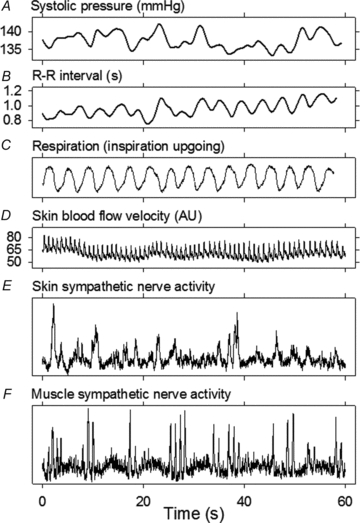
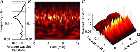
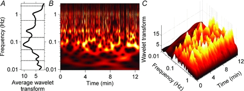
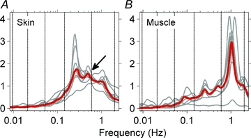
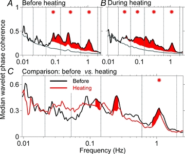
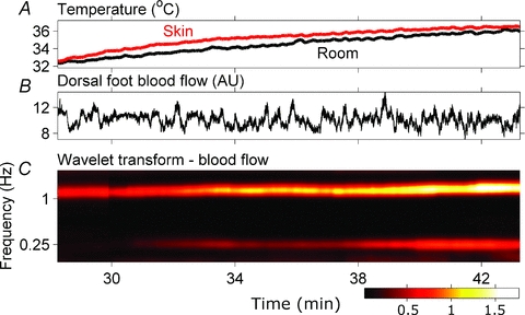
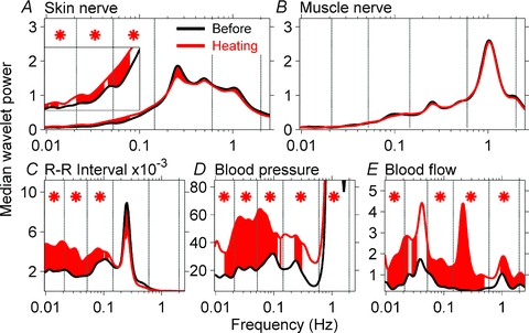
Frequency-domain analyses of simultaneously recorded skin and muscle sympathetic nerve activities may yield unique information on otherwise obscure central processes governing human neural outflows. We used wavelet transform and wavelet phase coherence methods to analyse integrated skin and muscle sympathetic nerve activities and haemodynamic fluctuations, recorded from nine healthy supine young men. We tested two null hypotheses: (1) that human skin and muscle sympathetic nerve activities oscillate congruently; and (2) that whole-body heating affects these neural outflows and their haemodynamic consequences in similar ways. Measurements included peroneal nerve skin and tibial nerve muscle sympathetic activities; the electrocardiogram; finger photoplethysmographic arterial pressure; respiration (controlled at 0.25 Hz, and registered with a nasal thermistor); and skin temperature, sweating, and laser-Doppler skin blood flow. We made recordings at ∼27°C, for ∼20 min, and then during room temperature increases to ∼38°C, over 35 min. We analysed data with a wavelet transform, using the Morlet mother wavelet and wavelet phase coherence, to determine the frequencies and coherences of oscillations over time. At 27°C, skin and muscle nerve activities oscillated coherently, at ever-changing frequencies between 0.01 and the cardiac frequency (∼1 Hz). Heating significantly augmented oscillations of skin sympathetic nerve activity and skin blood flow, arterial pressure, and R-R intervals, over a wide range of low frequencies, and modestly reduced coordination between skin and muscle sympathetic oscillations. These results suggest that human skin and muscle sympathetic motoneurones are similarly entrained by external influences, including those of arterial baroreceptors, respiration, and other less well-defined brainstem oscillators. Our study provides strong support for the existence of multiple, time-varying central sympathetic neural oscillators in human subjects.


Figures

Figure 1
Figure 1. Recording at room temperature from Subject 3
Respiration was controlled well by this and other subjects, who breathed with a metronome at 0.25 Hz.
Figure 2
Figure 2. Wavelet transform of skin sympathetic nerve activity from Subject 3
A, the average wavelet transform of skin sympathetic nerve activity for the recording period. B and C, a contour plot (B) and a three-dimensional plot (C) of the wavelet transform. The strongest periodicity, shown in yellow, is at the respiratory frequency (0.25 Hz). B and C illustrate the complexity of this subject's skin sympathetic rhythms, whose occurrence, frequency and strength were highly variable. The average data shown in A mask the major ongoing fluctuations present in the time series.
Figure 3
Figure 3. Wavelet transform of muscle sympathetic nerve activity from Subject 2
A, the average wavelet transform of the muscle sympathetic nerve activity. B and C, contour and three-dimensional plots of the same data. As with the analysis of skin sympathetic nerve activity (Fig. 2), the occurrence, frequency and strength of oscillations varied in major ways during the recording. This is particularly evident in the ∼0.1 Hz rhythm, which appeared and disappeared at highly variable rates. The average data shown in A do not accurately represent these ongoing fluctuations.
Figure 4
Figure 4. Median (red) and individual (grey) wavelet power spectra of skin and muscle sympathetic neurograms before heating
The strongest periodicity in skin sympathetic nerve activity (A) was at the respiratory frequency. A rhythm at the cardiac frequency was present, but was not prepossessing. The arrow points to the second harmonic of the respiratory frequency. The strongest periodicity in muscle sympathetic activity (B) was at the cardiac frequency. These median data also show oscillations at the respiratory frequency and ∼0.1 Hz.
Figure 5
Figure 5. Median (A) and individual phase coherences (B) between skin and muscle sympathetic nerve activities for all subjects
The heavy black lines indicate median (A) and individual (B) phase coherences; the grey lines indicate average surrogate values (see Methods), and the red areas indicate coherence which was significantly different (greater than 2 standard deviations above) from the surrogate values. The asterisks indicate frequency ranges within which skin and muscle coherences were significant. In all subjects, skin and muscle sympathetic nerve activities were significantly coherent over a broad range, including cardiac frequencies (extreme right of individual data panels). The group median data (A) obscure the significant narrow frequency ranges over which skin and muscle sympathetic nerve activities are coherent in individuals.
Figure 6
Figure 6. Median skin and muscle sympathetic activity wavelet phase coherence before and during heating
The black lines in A and B indicate median group coherences, the grey lines indicate average surrogate values, and the red shaded areas indicate coherences in 115 frequency ranges which are greater than 2 standard deviations above the surrogate values. Asterisks indicate conventional frequency ranges, in which the mean coherences are significantly above the mean surrogate values. C compares coherences before and during heating. Note that in C, the scale is narrower than in A and B. The only frequency range over which this reduction is significant is the cardiac frequency (*).
Figure 7
Figure 7. Skin blood flow responses of Subject 4 to heating
The blood flow signal (B) does not show obvious changes with heating in the time domain. The wavelet transform, which decomposes the signal into several oscillatory components and their variation in time documents a steady increase of the amplitudes of oscillatory components at cardiac and respiratory frequencies with heating (C).
Figure 8
Figure 8. Influence of heating on wavelet power spectra of neural and haemodynamic signals
Wavelet power spectra of signals are compared before and during heating. Red shaded areas indicate significant effect of heating at specific frequencies and asterisks indicate significances within the frequency ranges. Skin sympathetic nerve wavelet power increased significantly during heating at low frequencies (A, inset), but muscle sympathetic nerve wavelet power (B) was not affected by heating. R-R interval, blood pressure, and skin blood flow wavelet powers were significantly increased by heating, primarily at low frequencies.

References

    1. Akselrod S, Gordon D, Ubel FA, Shannon DC, Barger AC, Cohen RJ. Power spectrum analysis of heart rate fluctuation: a quantitative probe of beat-to-beat cardiovascular control. Science. 1981;213:220–222. - PubMed
    1. Badra LJ, Cooke WH, Hoag JB, Crossman AA, Kuusela TA, Tahvanainen KUO, Eckberg DL. Respiratory modulation of human autonomic rhythms. Am J Physiol Heart Circ Physiol. 2001;280:H2674–H2688. - PubMed
    1. Baldwin BA, Ingram DL. The influence of hypothalamic temperature and ambient temperature on thermoregulatory mechanisms in the pig. J Physiol. 1998;198:517–529. - PMC - PubMed
    1. Bandrivskyy A, Bernjak A, McClintock P, Stefanovska A. Wavelet phase coherence analysis: application to skin temperature and blood flow. Cardiovasc Eng. 2004;4:89–93.
    1. Barman SM, Gebber GL. Subgroups of rostral ventrolateral medullary and caudal medullary raphe neurons based on patterns of relationship to sympathetic nerve discharge and axonal projections. J Neurophysiol. 1997;77:65–75. - PubMed

Publication types

LinkOut - more resources