Case Reports
. 2012 Apr;9(4):590-7.
doi: 10.1016/j.hrthm.2011.11.006. Epub 2011 Nov 7.

Developmentally regulated SCN5A splice variant potentiates dysfunction of a novel mutation associated with severe fetal arrhythmia

Affiliations
Case Reports

Developmentally regulated SCN5A splice variant potentiates dysfunction of a novel mutation associated with severe fetal arrhythmia

Lisa L Murphy et al. Heart Rhythm. 2012 Apr.

Abstract

Background: Congenital long-QT syndrome (LQTS) may present during fetal development and can be life-threatening. The molecular mechanism for the unusual early onset of LQTS during fetal development is unknown.

Objective: We sought to elucidate the molecular basis for severe fetal LQTS presenting at 19 weeks' gestation, the earliest known presentation of this disease.

Methods: Fetal magnetocardiography was used to demonstrated torsades de pointes and a prolonged rate-corrected QT interval. In vitro electrophysiological studies were performed to determine functional consequences of a novel SCN5A mutation found in the fetus.

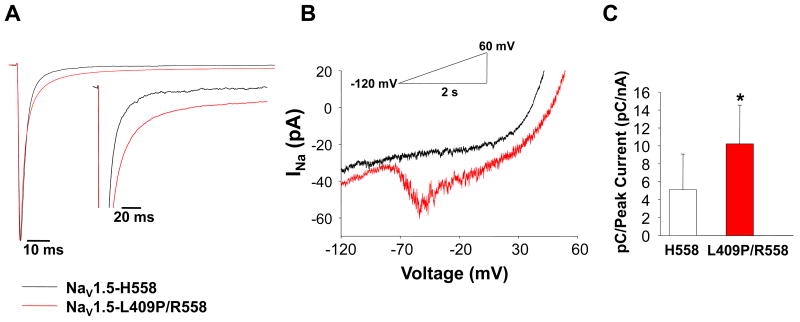
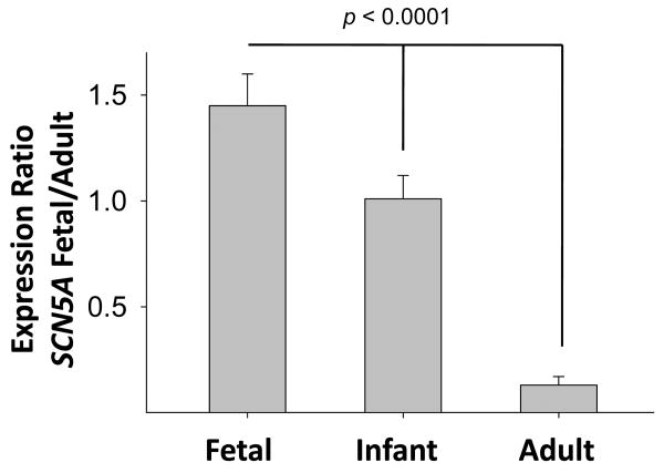
Results: The fetus presented with episodes of ventricular ectopy progressing to incessant ventricular tachycardia and hydrops fetalis. Genetic analysis disclosed a novel, de novo heterozygous mutation (L409P) and a homozygous common variant (R558 in SCN5A). In vitro electrophysiological studies demonstrated that the mutation in combination with R558 caused significant depolarized shifts in the voltage dependence of inactivation and activation, faster recovery from inactivation, and a 7-fold higher level of persistent current. When the mutation was engineered in a fetal-expressed SCN5A splice isoform, channel dysfunction was markedly potentiated. Also, R558 alone in the fetal splice isoform evoked a large persistent current, and hence both alleles were dysfunctional.

Conclusion: We report the earliest confirmed diagnosis of symptomatic LQTS and present evidence that mutant cardiac sodium channel dysfunction is potentiated by a developmentally regulated alternative splicing event in SCN5A. Our findings provide a plausible mechanism for the unusual severity and early onset of cardiac arrhythmia in fetal LQTS.


Conflict of interest statement

Conflicts of Interest: None

Figures

Figure 1
Figure 1. Fetal doppler echocardiogram and magnetocardiogram
(A) Pulsed wave Doppler of the fetal aorta at 20 weeks gestation. Normal conducted beats (red arrows) are interrupted by frequent premature beats (PBs), and 2-3 beat runs of tachycardia with variable cycle length (white arrows). The flow velocity is maintained during the short runs of tachycardia, suggesting stroke volume is only slightly diminished. (B) Signal averaged ‘butterfly plot’ determined by fetal magnetocardiography during sinus rhythm at 20-6/7 weeks gestation illustrating T-wave alternans, a QTc interval of 604 msec, normal PR interval and a QRS duration that was slightly prolonged for age (0.69 msec). (C) Representative rhythm trace obtained by fetal magnetocardiography at 20-6/7 weeks gestation. Arrows indicate sinus beats interrupted by two episodes of non-sustained polymorphic ventricular (V) tachycardia with varying cycle length (400 to 200 msec). (D) Pulsed wave Doppler of the middle cerebral artery at 22 weeks gestation illustrating fetal arrhythmia. At this time, the fetus was severely hydropic with very poor systolic function (not shown). The tracing shows the onset of a sustained period (>3500 msec) of ventricular tachycardia (white arrows), initiating by a premature beat (PB). During the tachycardia, there is decreased stroke volume as evidenced by the extremely low velocity Doppler signals. The tachycardia cycle length was 480 to 210 ms.
Figure 2
Figure 2. Biophysical properties of WT and mutant sodium channels
(A) Representative whole-cell current recordings from cells expressing either WT (Adult NaV1.5-H558) or mutant (Adult NaV1.5-L409P/R558) channels (voltage protocol shown as inset). (B) Current-voltage relationships for WT (n = 10) and mutant (n = 10) channels. Current was normalized to cell capacitance to give a measure of current density. (C) Superimposed curves representing the voltage dependence of steady-state inactivation (left y-axis) and conductance-voltage (right y-axis) relationships for WT and mutant channels. Lines represent average fits of the data with Boltzmann functions. (D) Time course of recovery from inactivation recorded using the illustrated voltage protocol. Biophysical fit parameters for all experiments are provided in Supplemental Table S1.
Figure 3
Figure 3. SCN5A-L409P/R558 exhibits increased persistent current
(A) Representative tetrodotoxin (TTX)-sensitive currents were normalized to the peak current measured at −30mV during a 200 ms depolarization to illustrate persistent current. The inset represents the same data plotted on an expanded vertical scale. Summary data are provided in Table 1. (B) WT and mutant currents elicited by voltage ramps defined by the inset. (C) Amount of charge moved between −70 and −30mV normalized to peak current and quantified. Charge movement was significantly (p<0.02) greater for mutant (10.2 ± 4.3 pC/nA, n = 9) than for WT (5.1 ± 4.0 pC/nA, n = 9) channels.
Figure 4
Figure 4. Developmental timing of SCN5A exon alternative splicing
Expression ratio (exon 6A / exon 6) of SCN5A mRNA transcripts expressed in human fetal (n = 4), infant (n = 4) and adult (n = 24) hearts determined by quantitative real-time RT-PCR. Differences among groups was significant at p<0.0001 (one-way ANOVA with Tukey test).
Figure 5
Figure 5. Functional consequences of R558 variant on fetal-NaV1.5
(A) Current density-voltage relationships recorded from cells expressing either WT (labeled Fetal-NaV1.5-H558; n = 11) or variant (Fetal NaV1.5-R558; n = 14) channels (voltage protocol same as in Fig. 2A). (B) Conductance-voltage relationships for WT and variant channels. (C) Steady-state voltage dependence of inactivation for WT (n = 10) and variant channels (n = 10). In (B) and (C) lines represent average fits of the data with Boltzmann functions. (D) Averaged TTX-sensitive persistent currents measured at −30mV during a 200 ms depolarization and normalized to peak current (n = 7). The inset represents the same data plotted on an expanded vertical scale. Biophysical fit parameters for all experiments are provided in Supplemental Table S1, and magnitude of persistent current is provided in Table 1.
Figure 6
Figure 6. Expression in fetal-NaV1.5 potentiates effect of SCN5A-L409P/R558
(A) Current density-voltage relationships comparing WT and mutant channels expressed in either adult or fetal NaV1.5 channels (voltage protocol same as in Fig. 2A; n = 10 for all groups). (B) Conductance-voltage relationships for WT and mutant channels. (C) Steady-state voltage dependence of inactivation for WT and mutant channels (n = 10-12). In (B) and (C) lines represent average fits of the data with Boltzmann functions. (D) Representative TTX-sensitive persistent currents measured at −30mV during a 200 ms depolarization and normalized to peak current. Biophysical fit parameters for all experiments are provided in Supplemental Table S1, and magnitude of persistent current is provided in Table 1.

Comment in

References

    1. Schwartz PJ, Crotti L. Long QT and short QT syndrome. In: Zipes DP, Jalife J, editors. Cardiac Electrophysiology: From Cell to Bedside. Fifth. Philadelphia: Elsevier/Saunders; 2009. pp. 731–744.
    1. Wedekind H, Smits JP, Schulze-Bahr E, et al. De novo mutation in the SCN5A gene associated with early onset of sudden infant death. Circulation. 2001;104:1158–1164. - PubMed
    1. Schwartz PJ, Priori SG, Dumaine R, et al. A molecular link between the sudden infant death syndrome and the long-QT syndrome. N Engl J Med. 2000;343:262–267. - PubMed
    1. Schulze-Bahr E, Fenge H, Etzrodt D, et al. Long QT syndrome and life threatening arrhythmia in a newborn: molecular diagnosis and treatment response. Heart. 2004;90:13–16. - PMC - PubMed
    1. Wang DW, Crotti L, Shimizu W, et al. Malignant perinatal variant of long-QT syndrome caused by a profoundly dysfunctional cardiac sodium channel. Circ Arrhythm Electrophysiol. 2008;1:370–378. - PMC - PubMed

Publication types

Substances