Review
. 2011 Nov 7;17(11):1381-90.
doi: 10.1038/nm.2514.

The neutrophil in vascular inflammation

Affiliations
Review

The neutrophil in vascular inflammation

Mia Phillipson et al. Nat Med. .

Abstract

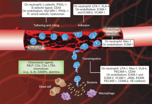
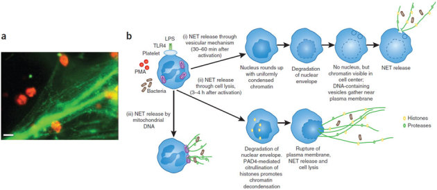
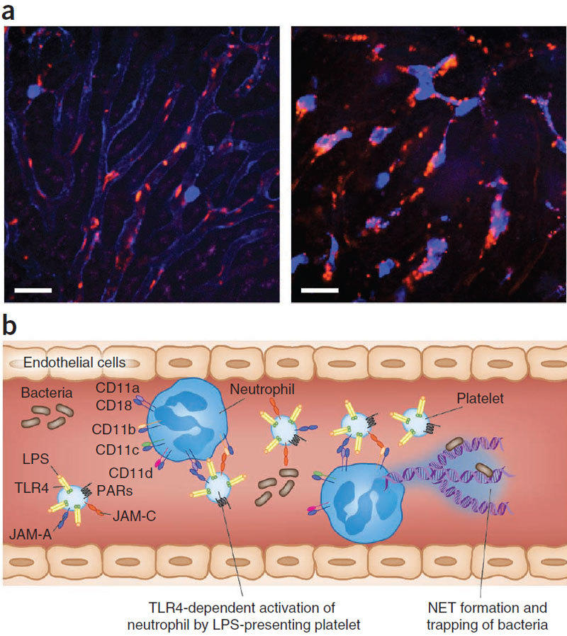
Here we focus on how neutrophils have a key regulatory role in vascular inflammation. Recent studies using advanced imaging techniques have yielded new insights into the mechanisms by which neutrophils contribute to defense against bacterial infections and also against sterile injury. In these settings, neutrophils are recruited by various mechanisms depending on the situation. We also describe how these processes may be disrupted in systemic infections, with a particular emphasis on mouse models of sepsis. Neutrophils are often immobilized in the lungs and liver during systemic infections, and this immobilization may be a mechanism through which bacteria can evade the innate immune response or allow neutrophils to form neutrophil extracellular traps that trap and kill bacteria in blood. The platelet is also an important player in sepsis, and we describe how it collaborates with neutrophils in the formation of neutrophil extracellular traps.


Conflict of interest statement

The authors declare no competing financial interests.

Figures

Figure 1
Figure 1. The neutrophil recruitment cascade.
Intravital confocal microscopy image of a cremasteric postcapillary venule with added cartoon cells illustrating the consecutive steps of the recruitment of circulating neutrophils to localized inflammation (infection or necrosis). The white boxes show the adhesion molecules involved in each step. Endothelial cell junctions are stained with monoclonal antibodies to CD31 (red). Endothelial upregulation of adhesion molecules results in interactions between selectins and their ligands on neutrophils, leading to neutrophil tethering and rolling. Chemokines sequestered on the luminal endothelium induce conformation changes of neutrophil β2 integrins, which results in neutrophil adhesion and crawling. Mechanotactic and chemotactic guidance signals direct crawling neutrophils to junctional transmigration sites closer to the source of chemotactic agent, examples of which are given in the green box.
Figure 2
Figure 2. Proposed model of PTEN, PI3K and p38 MAPK function during neutrophil chemotaxis.
(a) The localizations of PIP3 (red), PTEN and PIP2 (green) are shown in a neutrophil migrating toward an intermediary chemoattractant (CXCR2 ligand (CXCL2–MIP-2 or CXCL8-IL-8)). LPS inhibits IL-8–induced chemotaxis. (b) The localizations of PIP3 (red), PTEN and PIP2 (green) are shown in a cell migrating toward an end-target chemoattractant (formylpeptide). p38 MAPK mediates both PTEN localization (green arrows) as well as chemotaxis (red dotted arrow) in response to end-target chemoattractants. LPS inhibits p38 MAPK and thereby inhibits chemotaxis. (c) The localizations of PTEN and PIP2 (green) are shown in a neutrophil migrating in opposing gradients of end-target (formylpeptide) and intermediary (CXCR2 ligand) chemoattractants. Because PTEN antagonizes any PIP3 accumulation, all chemotaxis occurs through p38 MAPK (red dotted arrow). LPS stops chemotaxis by overriding the effects of the chemoattractants through p38 MAPK inhibition.
Figure 3
Figure 3. Neutrophils move to sites of sterile injury by intravascular crawling.
(a) Time-lapse images using spinning-disk confocal intravital microscopy show the response of neutrophils (green) to focal hepatic necrosis (red, propiodium iodide). Scale bar, 200 μm (ref. 37). (b) Images of neutrophils homing (green) to sterile injury (red, propiodium iodide) in the liver of untreated mice (left) or after treatment with the fMLP receptor inhibitor cyclosporine H (right). (c) Neutrophils within the liver vasculature can prioritize an fMLP gradient when they are subjected to competing gradients of CXCL2 and fMLP, thus allowing them to chemotax toward the site of focal necrosis.
Figure 4
Figure 4. Formation of neutrophil extracellular traps.
(a) LPS-induced platelet adhesion to neutrophils resulted in NET formation. Representative image of neutrophils visualized by white light through an orange filter using dark-field illumination and fluorescence microscopy with SYTOX Green to stain extracellular DNA green. Scale bar, 10 μm. (b) Various mechanisms of NET release have been observed. (i) NETs can be released through a vesicular mechanism,. Initially, the neutrophils become rounded with uniformly condensed chromatin and then undergo nuclear envelope breakdown. Within these cells, small vesicles containing DNA can be seen in the cytoplasm near the plasma membrane. The DNA-containing vesicles eventually fuse with the plasma membrane, and NETs are released to trap bacteria. (ii) NETs can also be released through cell lysis, and this typically takes longer than the vesicular-mediated mechanism,,,. The nuclear envelope is degraded, and chromatin decondensation occurs because of PAD4-mediated citrullination of histones. (iii) NET release by mitochondria has also been observed, in one study, although the steps of this process remain poorly characterized.
Figure 5
Figure 5. Platelet-neutrophil interactions within the liver vasculature during endotoxemia.
(a) Intravital spinning-disk confocal visualization of neutrophils (blue) and platelets (red) in liver sinusoids in healthy (left) and endotoxemic (right) mice. Although no neutrophil-platelet interactions were detected in healthy mice (left), a notable difference was observed in endotoxemic mice, in which neutrophil-platelet interactions were very common within the liver vasculature (colocalization of red and blue, right). Scale bar, 25 μm. (b) Schematic showing the various cell-surface molecules on platelets and neutrophils. TLR4 expression by the platelet has been shown to be important for neutrophil activation in response to LPS. JAM-A and JAM-C expressed by platelets interact with neutrophil integrins CD11a-CD18 and CD11b-CD18, respectively. Interactions between platelets can also stimulate NET release from neutrophils.

References

    1. Auffray C, et al. Monitoring of blood vessels and tissues by a population of monocytes with patrolling behavior. Science. 2007;317:666–670. - PubMed
    1. Ley K, Laudanna C, Cybulsky MI, Nourshargh S. Getting to the site of inflammation: the leukocyte adhesion cascade updated. Nat. Rev. Immunol. 2007;7:678–689. - PubMed
    1. Petri B, Phillipson M, Kubes P. The physiology of leukocyte recruitment: an in vivo perspective. J. Immunol. 2008;180:6439–6446. - PubMed
    1. Luo BH, Carman CV, Springer TA. Structural basis of integrin regulation and signaling. Annu. Rev. Immunol. 2007;25:619–647. - PMC - PubMed
    1. Phillipson M, et al. Intraluminal crawling of neutrophils to emigration sites: a molecularly distinct process from adhesion in the recruitment cascade. J. Exp. Med. 2006;203:2569–2575. - PMC - PubMed

Publication types

MeSH terms

Substances