

Disparate effects of depletion of CD1d-reactive T cells during early versus late stages of disease in a genetically susceptible model of lupus
- PMID: 22065098
- PMCID: PMC3412757
- DOI: 10.1177/0961203311428459
Disparate effects of depletion of CD1d-reactive T cells during early versus late stages of disease in a genetically susceptible model of lupus
Abstract
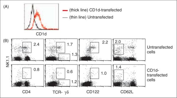
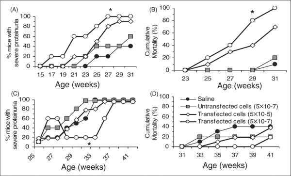
Some T cells react with lipid antigens bound to antigen-presenting molecule CD1d. Numbers and functions of a subset of such lipid-reactive T cells are reduced in patients with systemic lupus erythematosus (SLE) and their relatives, as well as in genetically susceptible and chemically induced animal models of lupus-like disease. We have reported that the germline deletion of CD1d exacerbates lupus, suggesting a protective role of these cells in the development of lupus. The use of a knockout mouse model in this study, however, did not allow examination of the role of these cells at different stages of disease. Here, we describe an approach to deplete CD1d-dependent T cells, which allowed us to investigate the role of these cells at different stages of disease in genetically lupus-prone NZB/NZW F1 (BWF1) mice. Repeated intravenous injections of large numbers of CD1d-transfected cells resulted in ∼50-75% reduction in these cells, as defined by the expression of CD4, NK1.1 and CD122, and lack of expression of CD62 ligand. TCR γδ (+)NK1.1(+) cells were also reduced in the recipients of CD1d-transfected cells as compared with control recipients. Such depletion of CD1d-reactive T cells in preclinical BWF1 mice resulted in disease acceleration with a significant increase in proteinuria and mortality. In older BWF1 mice having advanced nephritis, however, such depletion of CD1d-reactive T cells resulted in some disease improvement. Taken together, these data as well as our published studies suggest that CD1d-reactive T cells protect against the development of lupus in animal models. However, these cells appear to be unable to suppress established lupus nephritis in these animals, and might even play a disease aggravating role in late stages of disease.
Figures
References
-
- Porcelli SA, Modlin RL. The CD1 system: antigen-presenting molecules for T cell recognition of lipids and glycolipids. Annu Rev Immunol. 1999;17:297–329. - PubMed
-
- De Silva AD, Park JJ, Matsuki N, et al. Lipid protein interactions: the assembly of CD1d1 with cellular phospholipids occurs in the endoplasmic reticulum. J Immunol. 2002;168:723–733. - PubMed
-
- Gumperz JE, Roy C, Makowska A, et al. Murine CD1d-restricted T cell recognition of cellular lipids. Immunity. 2000;12:211–221. - PubMed
-
- Cho YN, Kee SJ, Lee SJ, et al. Numerical and functional deficiencies of natural killer T cells in systemic lupus erythematosus: their deficiency related to disease activity. Rheumatology (Oxford) 2011;50:1054–1063. - PubMed
Publication types
MeSH terms
Substances
Grants and funding
LinkOut - more resources
Full Text Sources
Medical
Research Materials
