. 2012 Jan 5;119(1):180-7.
doi: 10.1182/blood-2011-08-374561. Epub 2011 Nov 7.

Tcl1 interacts with Atm and enhances NF-κB activation in hematologic malignancies

Affiliations

Tcl1 interacts with Atm and enhances NF-κB activation in hematologic malignancies

Eugenio Gaudio et al. Blood. .

Abstract

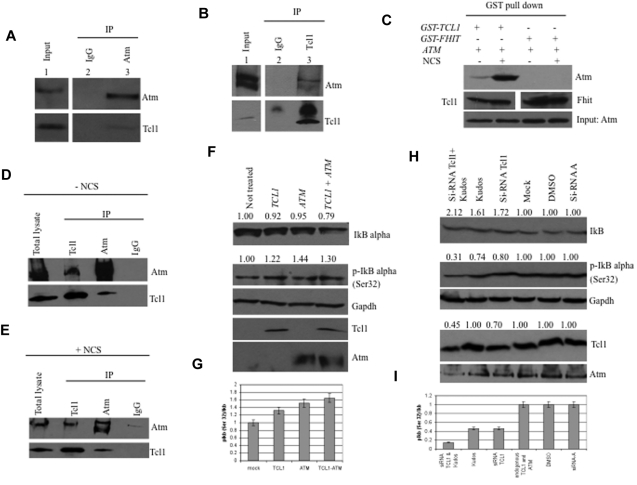
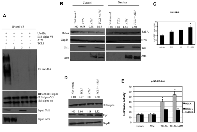
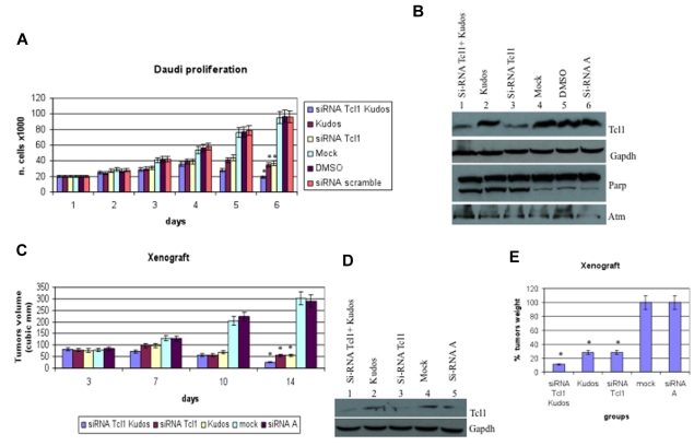
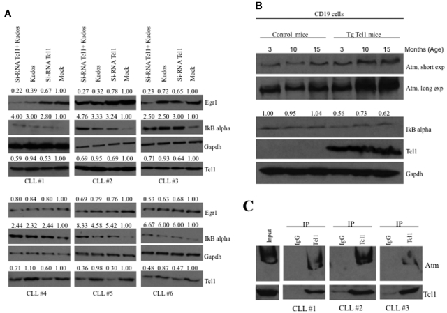
The T-cell leukemia/lymphoma 1 (TCL1) oncogene is a target of chromosomal translocations and inversions at 14q31.2, and its rearrangement in T cells causes T-cell prolymphocytic leukemias. TCL1 dysregulation in B cells is responsible for the development of an aggressive form of chronic lymphocytic leukemia (CLL), the most common human leukemia. We have investigated the mechanisms underlying the oncogenic functions of Tcl1 protein using a mass spectrometry approach and have identified Atm (ataxia-telangiectasia mutated) as a candidate Tcl1-interacting protein. The Tcl1-Atm complex formation was validated by coimmunoprecipitation experiments. Importantly, we show that the association of Atm with Tcl1 leads to enhanced IκBα phosphorylation and ubiquitination and subsequent activation of the NF-κB pathway. Our findings reveal functional cross-talk between Atm and Tcl1 and provide evidence for a novel pathway that could be targeted in leukemias and lymphomas.


Figures

Figure 1
Figure 1
Tcl1 interacts with Atm, and both affect IκBα expression. (A) HEK293 cells were stably transfected with expression plasmid encoding FLAG-ATM and then infected with Ad-TCL1 wild-type (MOI 100). Forty-eight hours after infection, whole-cell lysates were immunoprecipitated with anti-Atm (resin conjugated to Atm). The immunoprecipitates were analyzed by Western blot with anti-Atm or anti-Tcl1. Input: lysate expression of Atm (top) and Tcl1 (bottom). (B) HEK293 cells were transiently cotransfected with expression plasmids encoding mammalian FLAG-ATM (8 μg) and wild-type TCL1 (6 μg). Forty-eight hours after transfection, cell lysates were immunoprecipitated with anti-TCL1. The immunoprecipitates were analyzed by Western blot with anti-Atm or anti-Tcl1. Input: lysate expression of Atm (top) and Tcl1 (bottom). (C) HEK293 cells were transiently cotransfected with expression plasmids encoding mammalian Omni-GST-TCL1 (6 μg) or Omni-GST-FHIT (6 μg) and FLAG-HIS-ATM (8 μg). Forty-eight hours after transfection, cells were treated with neocarzinostatin (NCS) 200 ng/mL for 2 hours. Cell lysates were GST-pulled down and immunoblotted with anti-His6, anti-Tcl1, and anti-Fhit. Input: lysate expression of Atm. (D-E) Daudi cells were untreated or treated with hydroxyurea, 50mM for 3 hours, then lysed and immunoprecipitated, performed with anti-Atm, anti-Tcl1, or IgG. The precipitates were analyzed by Western blot with anti-Atm or anti-Tcl1. (F) Tcl1 and Atm coexpression is associated with decreased IκBα total protein. HEK293 cells were mock-transfected or transfected with TCL1 or ATM plasmids separately or together (4 μg for each plasmid, including empty vector). Forty-eight hours later, cells were treated with hydroxyurea 50mM for 3 hours and then analyzed by Western blot with the indicated antibodies. (G) Tcl1 and Atm coexpression is associated with increased IκBα (Ser32) phosphorylation as shown in the graph. (H) Daudi cells were mock-transfected or transfected with si-TCL1 or si-Scr, and then untreated or treated with Kudos 55933 or DMSO. The level of IκBα was measured by Western blot. (I) IκBα (Ser 32) levels are shown in the graph. Data are representative of 3 independent experiments.
Figure 2
Figure 2
Expression of Tcl1 and Atm activates the NF-κB pathway. (A) HEK293 cells were transfected with indicated plasmids. Forty-eight hours later, cells were treated with MG132, 10μM for 3 hours, and then lysates immunoprecipitated with anti-V5. The precipitates were analyzed by Western blot with anti-HA or anti-IκBα. Input: lysate expression of Tcl1 and Atm. (B) HEK293 cells were mock-transfected or transfected with TCL1 and ATM plasmids separately or together (4 μg each plasmid). Forty-eight hours later, cells were treated with hydroxyurea for 3 hours, fractionated into cytosolic and nuclear fractions, and analyzed by Western blot with the indicated antibodies. (C) mRNA levels of EGR1 were measured by quantitative RT-PCR. HEK293 cells were mock-transfected or transfected with TCL1 and ATM plasmids, separately or together for 48 hours, and then treated with hydroxyurea, 50mM for 3 hours). Fold changes of EGR1 were calculated using the 2−ΔCt method. GAPDH mRNA levels were used as an internal normalization control. Samples transfected with TCL1 and ATM have been normalized to mock-transfected sample. (D) HEK293 cells were transfected with TCL1 and ATM (4 μg for each plasmid), treated with hydroxyurea for 3 hours and with MG132, 10μM 6 hours, and protein lysates run on gel. (E) Tcl1 activates NF-κB–dependent transcription synergistically with Atm. HEK-293 cells were cotransfected with 50 ng of pNF-κB-Luc reporter and 50 ng of pRL-TK Renilla reporter constructs. In addition, 0.75 μg of CMV5-empty vector, 0.75 μg of pcDNA 3.1 empty vector, 0.75 μg of CMV5-TCL1 WT, and 0.75 μg of pcDNA 3.1 empty vector constructs were used; 5 ng of pFC-MEKK was added where indicated. Cells were treated with 10μM Kudos for 5 hours, where indicated. Data are representative of 3 independent experiments. (C,E) Data are mean ± SEM of 3 independent experiments, and each is measured in triplicate. *P < .05.
Figure 3
Figure 3
Tcl1 and Atm are crucial for growth and proliferation of lymphoma cells in vitro and in vivo. (A) Daudi cells were mock-transfected or transfected with si-TCL1 or si-Scr and then untreated or treated with Kudos 55933 or DMSO. Cells were counted at 24-hour intervals. Results represent the average of 3 independent experiments. (B) Total lysates collected were analyzed by Western blot and tested with indicated antibodies. (C) Graph representing tumor volumes at indicated days during the experiment for the 5 groups indicated (4 mice/group). Tumor size was measured daily until the tumor reached 50 mm3. Then, 5 μg of synthetic si-TCL1or si-Scr diluted in Lipofectamine and with or without Kudos (50 μL total volume) were injected directly into the tumors and at 3, 7, and 10 days. Tumors were measured on the day of the injections and 4 days after the last injection. (D) Total lysates from tumors were analyzed by Western blot and tested with indicated antibodies. (E) Xenograft tumors were weighed. Data are mean ± SEM of 3 independent experiments, and each is measured in triplicate. *P < .05.
Figure 4
Figure 4
Tcl1 and Atm affect the NF-κB pathway in CLL patient samples and Tcl1 transgenic mice. (A) B-CLL samples were transfected with si-TCL1 for 36 hours or treated with Kudos (10μM, 12 hours) or both and in all cases treated with hydroxyurea, 50mM 3 hours. IκBα and Egr1 expression was detected by Western blot. (B) B-cell CD19 isolated from mouse spleen was lysed and analyzed by Western blot with indicated antibodies. Spleens were excised from wild-type and Tcl1 transgenic mice at 3, 10, and 15 months of age. Data are representative of 3 independent experiments. (C) Whole B-CLL cell lysates were immunoprecipitated with anti-Tcl1 antibody. The immunoprecipitates were analyzed by immunoblotting (IB) with anti-Atm and anti-Tcl1 antibodies. Input: total lysate.

References

    1. Pekarsky Y, Halas C, Isobe M, Russo G, Croce CM. Abnormalities at 14q32.1 in T cell malignancies involve two oncogenes. Proc Natl Acad Sci U S A. 1999;96(6):2949–2951. - PMC - PubMed
    1. Narducci MG, Fiorenza MT, Kang SM, et al. TCL1 participates in early embryonic development and is overexpressed in human seminomas. Proc Natl Acad Sci U S A. 2002;99(18):11712–11717. - PMC - PubMed
    1. Virgilio L, Narducci MG, Isobe M, et al. Identification of the TCL1 gene involved in T-cell malignancies. Proc Natl Acad Sci U S A. 1994;91(26):12530–12534. - PMC - PubMed
    1. Madani A, Choukroun V, Soulier J, et al. Expression of p13MTCP1 is restricted to mature T-cell proliferations with t(X;14) translocations. Blood. 1996;87(5):1923–1927. - PubMed
    1. Narducci MG, Virgilio L, Engiles JB, et al. The murine Tcl1 oncogene: embryonic and lymphoid cell expression. Oncogene. 1997;15(8):919–926. - PubMed

Publication types

MeSH terms