. 2011 Nov 14;195(4):617-29.
doi: 10.1083/jcb.201106037. Epub 2011 Nov 7.

Hsp42 is required for sequestration of protein aggregates into deposition sites in Saccharomyces cerevisiae

Affiliations

Hsp42 is required for sequestration of protein aggregates into deposition sites in Saccharomyces cerevisiae

Sebastian Specht et al. J Cell Biol. .

Abstract

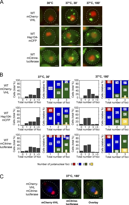
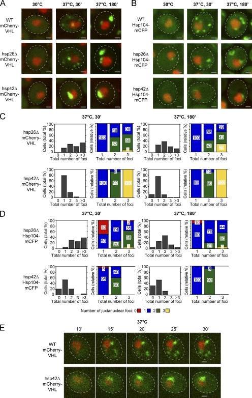
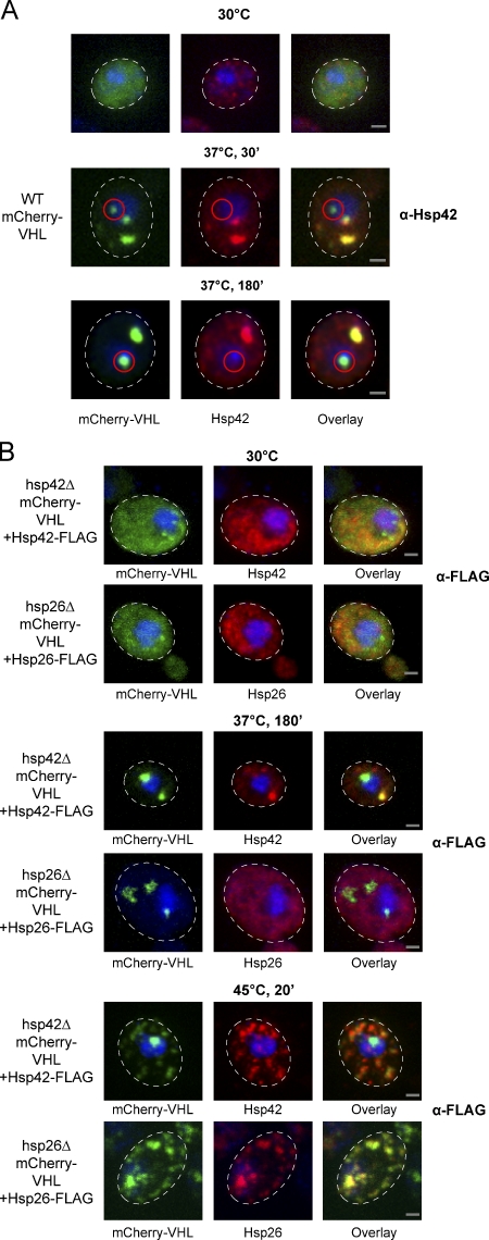
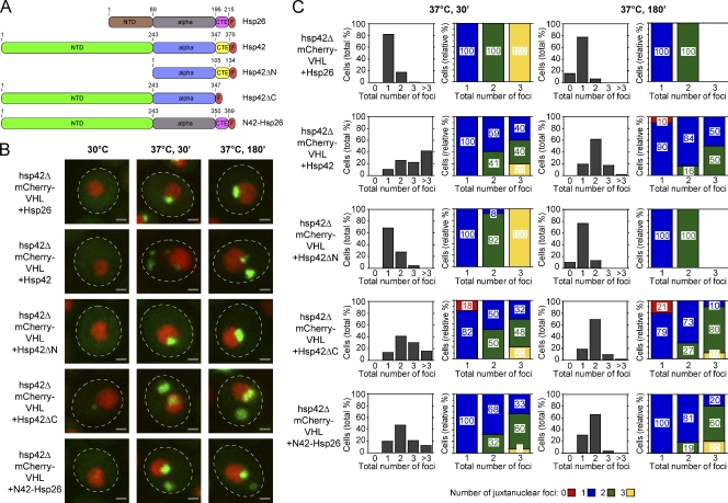
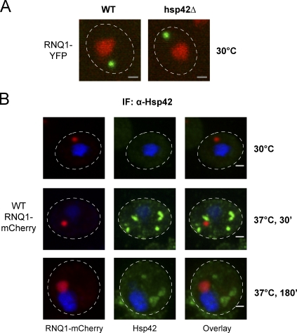
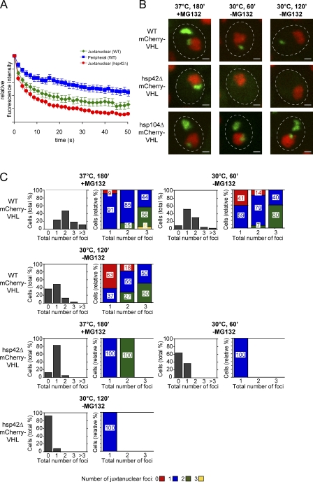
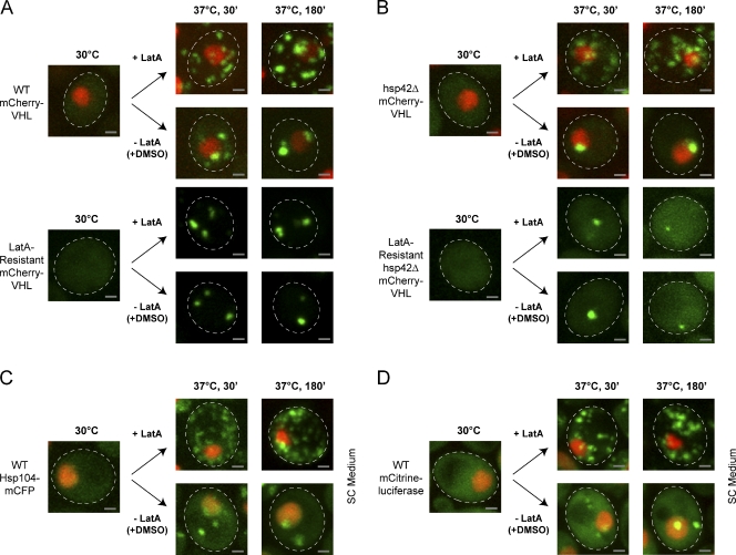
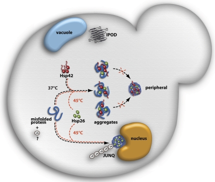
The aggregation of proteins inside cells is an organized process with cytoprotective function. In Saccharomyces cerevisiae, aggregating proteins are spatially sequestered to either juxtanuclear or peripheral sites, which target distinct quality control pathways for refolding and degradation. The cellular machinery driving the sequestration of misfolded proteins to these sites is unknown. In this paper, we show that one of the two small heat shock proteins of yeast, Hsp42, is essential for the formation of peripheral aggregates during physiological heat stress. Hsp42 preferentially localizes to peripheral aggregates but is largely absent from juxtanuclear aggregates, which still form in hsp42Δ cells. Transferring the amino-terminal domain of Hsp42 to Hsp26, which does not participate in aggregate sorting, enables Hsp26 to replace Hsp42 function. Our data suggest that Hsp42 acts via its amino-terminal domain to coaggregate with misfolded proteins and perhaps link such complexes to further sorting factors.


Figures

Figure 1.
Figure 1.
Spatiotemporal organization of protein aggregates in yeast cells. (A) Time-dependent changes in the localization of mCherry-VHL, Hsp104-mCFP, and mCitrine-luciferase (all green) in WT cells grown at 30°C and after a shift to 37°C for 30 and 180 min. MG132 was added before the temperature shift. Nuclei were visualized by coexpressing HTB1-cerulean or HTB1-mCherry (red). (B) Number (shaded bars) and localization (colored bars) of mCherry-VHL, Hsp104-mCFP, and mCitrine-luciferase aggregates in WT cells after incubation at 37°C for 30 and 180 min. The total number of foci per cell (juxtanuclear and peripheral) is depicted in all diagrams on the x axis. Quantifications are based on the analysis of 50–120 cells and are representatives of multiple experimental repeats. (C) Different substrates are sorted to the same compartments. mCherry-VHL (red) and mCitrine-luciferase (green) were coexpressed in WT cells. Protein localizations were determined after a temperature shift to 37°C for 180 min, revealing colocalization of mCherry-VHL and mCitrine-luciferase. Nuclei were visualized by coexpressing HTB1-cerulean (blue). The dashed lines indicate the border of respective yeast cells. Bars, 1 µm.
Figure 2.
Figure 2.
Hsp42 is essential for the targeting of misfolded proteins to peripheral aggregates. (A and B) Time-dependent changes in the localization of mCherry-VHL (A) and Hsp104-mCFP (B; both green) at 30°C and after a shift to 37°C for 30 and 180 min in the isogenic WT, hsp26Δ, and hsp42Δ strains. MG132 was added before the temperature shift. Nuclei were visualized by coexpressing HTB1-cerulean or HTB1-mCherry (red). (C and D) Number (shaded bars) and localization (colored bars) of mCherry-VHL (C) and Hsp104-mCFP (D) aggregates in hsp26Δ and hsp42Δ cells after incubation at 37°C for 30 and 180 min. MG132 was added before the temperature shift. The total number of foci per cell is depicted on the x axis in all diagrams. Quantifications are based on the analysis of 70–90 cells and are representatives of at least two experimental repeats. (E) In hsp42Δ cells, mCherry-VHL foci form exclusively at the nucleus. Time-lapse microscopy pictures of single WT and hsp42Δ cells expressing mCherry-VHL (green) after a shift to 37°C for the indicated time period. MG132 was added before the temperature shift. Nuclei were visualized by coexpressing HTB1-cerulean (red). The dashed lines indicate the border of respective yeast cells. Bars, 1 µm.
Figure 3.
Figure 3.
Hsp42 localizes exclusively to peripheral mCherry-VHL inclusions. (A) WT cells expressing mCherry-VHL were grown at 30°C and shifted to 37°C (+MG132). The cellular localizations of mCherry-VHL (green) and Hsp42 (red) were determined at the indicated time points. Hsp42 localization was determined by immunofluorescence using Hsp42-specific antibodies. Nuclei were visualized by coexpressing HTB1-cerulean (blue). Juxtanuclear mCherry-VHL foci that do not colocalize with Hsp42 are highlighted by red circles. (B) hsp42Δ or hsp26Δ cells coexpressing mCherry-VHL and Hsp42-FLAG or Hsp26-FLAG as indicated were grown at 30°C and shifted to 37°C (+MG132) for 180 min or to 45°C (+MG132) for 20 min. mCherry-VHL is depicted in green, and sHSP stainings using FLAG antibodies are shown in red. Nuclei were visualized by DAPI staining (blue). The dashed lines indicate the border of respective yeast cells. Bars, 1 µm.
Figure 4.
Figure 4.
The NTD of Hsp42 mediates sorting of aggregated proteins to peripheral inclusions. (A) Domain organization of Hsp26, Hsp42, and their variants. Both sHSPs consist of an NTD, a conserved α-crystallin domain (alpha), and a CTE. All constructs were C-terminally fused to a FLAG tag (F). Domain boundaries are indicated by residue numbers. All constructs were placed under control of the native HSP42 promoter and integrated at the Hsp42 locus in hsp42Δ cells. (B) hsp42Δ cells expressing mCherry-VHL and the indicated sHSP constructs were grown at 30°C and shifted to 37°C (+MG132). mCherry-VHL localization (green) was determined at the indicated time points. Nuclei were visualized by coexpressing HTB1-cerulean (red). The dashed lines indicate the border of respective yeast cells. Bars, 1 µm. (C) Number (shaded bars) and localization (colored bars) of mCherry-VHL inclusions in hsp42Δ cells expressing the indicated sHSP construct after incubation at 37°C for 30 and 180 min. MG132 was added before the temperature shift. The total number of foci per cell is shown on the x axis in all diagrams. Quantifications are based on the analysis of 40–130 cells and are representatives of two experimental repeats.
Figure 5.
Figure 5.
Hsp42 does not affect the localization of amyloidogenic aggregates. (A) RNQ1-YFP (green) was expressed in WT and hsp42Δ cells. Nuclei were visualized by coexpressing HTB1-mCherry (red). (B) WT cells expressing RNQ1-mCherry (green) were incubated at 30°C and shifted to 37°C for 30 min or 180 min (+MG132). The localization of Hsp42 (red) was determined by immunofluorescence (IF) using Hsp42-specific antibodies. Nuclei were visualized by DAPI staining (blue). The dashed lines indicate the border of respective yeast cells. Bars, 1 µm.
Figure 6.
Figure 6.
The juxtanuclear aggregates of hsp42Δ cells display altered protein dynamics and stability. (A) Juxtanuclear aggregates of hsp42Δ cells exhibit an increased exchange rate with the cytosolic mCherry-VHL pool. FLIP measurements of mCherry-VHL were performed in WT, hsp42Δ, and hsp104Δ cells after incubation at 37°C for 180 min (+MG132). Bleaching curves were calculated based on the analysis of 25 cells and the corresponding SEM. (B) mCherry-VHL aggregation foci are more rapidly resolved in the hsp42Δ strain, and their disintegration requires Hsp104-mediated protein disaggregation. WT, hsp42Δ, and hsp104Δ cells expressing mCherry-VHL (green) were grown at 30°C and shifted to 37°C for 180 min (+MG132). MG132 was washed out, and cells were shifted to 30°C for 120 min. De novo synthesis of mCherry-VHL was inhibited by the addition of 10 µg/ml cycloheximide. Nuclei were visualized by coexpressing HTB1-cerulean (red). The dashed lines indicate the border of respective yeast cells. Bars, 1 µm. (C) Number (shaded bars) and localization (colored bars) of mCherry-VHL aggregates are shown in the respective strain at the indicated time points. The total number of foci per cell is depicted in all diagrams on the x axis. Quantifications are based on the analysis of 60–80 cells and are representatives of multiple experimental repeats.
Figure 7.
Figure 7.
The actin cytoskeleton is required for aggregate compartmentalization. Reduction of mCherry-VHL (green) foci numbers during prolonged protein folding stress requires actin polymerization. Cells expressing mCherry-VHL were grown at 30°C and shifted to 37°C (+MG132). LatA or the same volume of DMSO as a control was added to the cells before a temperature shift. (A) mCherry-VHL localization was monitored in WT cells and cells containing a mutation in actin-1, which renders the actin cytoskeleton resistant to LatA. (B–D) hsp42Δ cells with or without the actin-1 mutation expressing mCherry-VHL (B) and WT cells expressing Hsp104-mCFP (C) or mCitrine-luciferase (D) were treated as described in A. All fluorescent reporter proteins are shown in green, and nuclei were visualized by coexpressing HTB1-cerulean (red). SC, synthetic complete. The dashed lines indicate the border of respective yeast cells. Bars, 1 µm.
Figure 8.
Figure 8.
Model depicting the roles of Hsp42 and Hsp26 in aggregate sorting in yeast cells experiencing sublethal folding stress. Hsp42 exclusively associates with peripheral protein aggregates during physiological folding stress (37°C) but is absent from juxtanuclear aggregates (JUNQ). The formation of peripheral foci depends on the elongated NTD of Hsp42. Hsp26 is absent from protein aggregates at 37°C but associates with peripheral and juxtanuclear aggregates during lethal heat stress (45°C). Hsp42 does not associate with the IPOD compartment harboring amyloidogenic proteins close to the vacuole. Ubiquitination (Ub) might serve as a sorting signal for juxtanuclear aggregates.

Similar articles

Cited by

References

    1. Abelson J.N., Guthrie C., Fink G.R. 2003. Guide to Yeast Genetics and Molecular Biology. Academic Press, New York: 933 pp
    1. Arrigo A.P. 2000. sHsp as novel regulators of programmed cell death and tumorigenicity. Pathol. Biol. (Paris). 48:280–288 - PubMed
    1. Ayscough K.R., Stryker J., Pokala N., Sanders M., Crews P., Drubin D.G. 1997. High rates of actin filament turnover in budding yeast and roles for actin in establishment and maintenance of cell polarity revealed using the actin inhibitor latrunculin-A. J. Cell Biol. 137:399–416 10.1083/jcb.137.2.399 - DOI - PMC - PubMed
    1. Basha E., Friedrich K.L., Vierling E. 2006. The N-terminal arm of small heat shock proteins is important for both chaperone activity and substrate specificity. J. Biol. Chem. 281:39943–39952 10.1074/jbc.M607677200 - DOI - PubMed
    1. Cashikar A.G., Duennwald M., Lindquist S.L. 2005. A chaperone pathway in protein disaggregation. Hsp26 alters the nature of protein aggregates to facilitate reactivation by Hsp104. J. Biol. Chem. 280:23869–23875 10.1074/jbc.M502854200 - DOI - PMC - PubMed

Publication types

MeSH terms

Substances