. 2012 Jan 1;72(1):201-9.
doi: 10.1158/0008-5472.CAN-11-1354. Epub 2011 Nov 7.

Use of multifunctional sigma-2 receptor ligand conjugates to trigger cancer-selective cell death signaling

Affiliations

Use of multifunctional sigma-2 receptor ligand conjugates to trigger cancer-selective cell death signaling

Dirk Spitzer et al. Cancer Res. .

Abstract

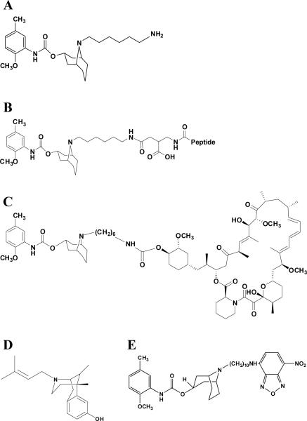
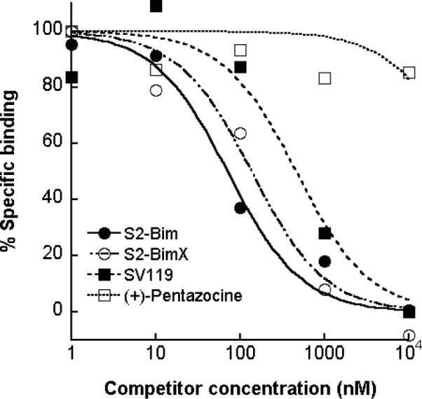
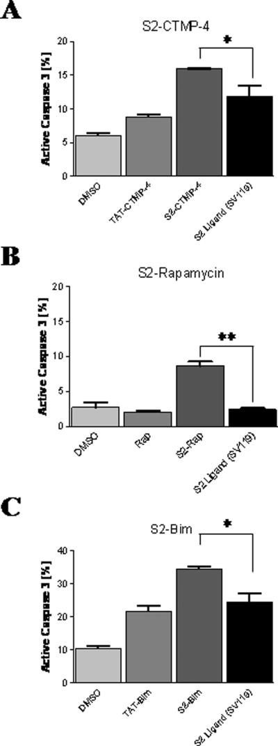
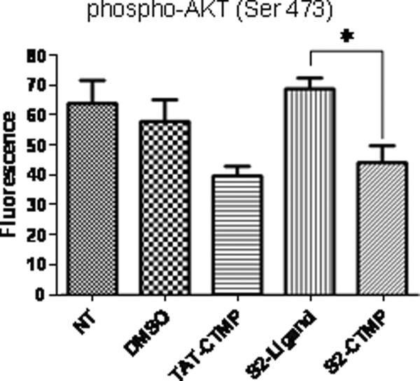
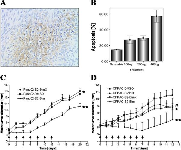
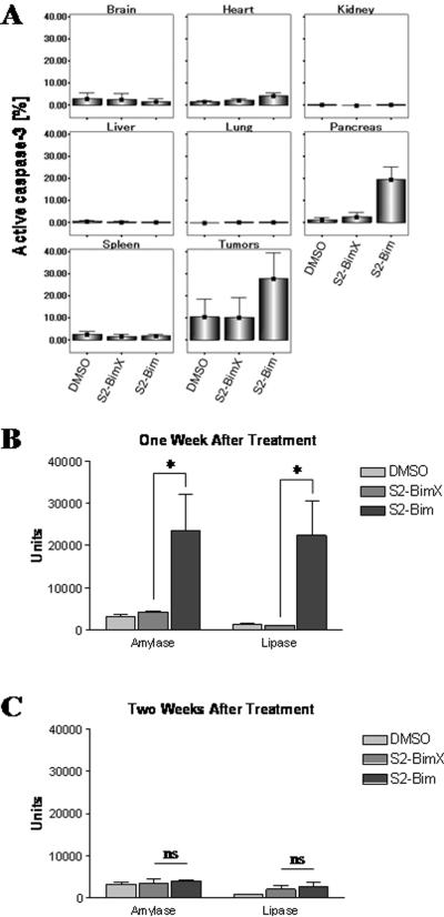
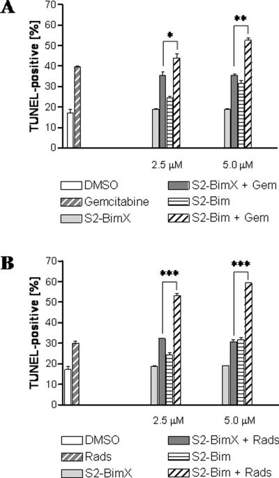
One major challenge in the development of cancer therapeutics is the selective delivery of the drugs to their cellular targets. In the case of pancreatic cancer, the σ-2 receptor is a unique target that triggers apoptosis upon activation. We have previously developed a series of chemical compounds with high affinity for the σ-2 receptor and showed rapid internalization of the ligands. One particular specific ligand of the σ-2 receptor, SV119, binds to pancreatic cancer cells and induces target cell death in vitro and in vivo. In this study, we characterized the ability of SV119 to selectively deliver other death-inducing cargos to augment the cytotoxic properties of SV119 itself. When conjugated to SV119, small molecules that are known to interfere with intracellular prosurvival pathways retained their ability to induce cell death, the efficiency of which was enhanced by the combinatorial effect of SV119 delivered with its small molecule cargo. Our findings define a simple platform technology to increase the tumor-selective delivery of small molecule therapeutics via σ-2 ligands, permitting chemotherapeutic synergy that can optimize efficacy and patient benefit.


Figures

Figure 1
Figure 1. Chemical structures of the relevant drugs used in this study
The parental compound SV119 (A) served as the basic structure for the generation of other conjugates. The basic structural organization of peptide-linked SV119 is shown in (B). The peptide sequences were as follows: S2-Bim (EIWIAQELRRIGDEFNAYYAR-OH), the inactive form S2-BimX (EIWIAQEARRIGAEFNAYYAR-OH), S2-CTMP-4 (LDPKLMKEEQMSQAQLFTRSFDDGL-OH), and the inactive derivative S2-CTMP-4X (LDPKLMKEEQMSQAQLATRSADDGL-OH). The conversion to functionally inactive peptide variants was done by Alanine substitution and is underlined. (C) The SV119-based conjugate S2-rapamycin. The sigma-1-specific ligand (+)-pentazocine (D) served as a negative control for binding studies in which the fluorescently-labeled sigma-2 ligand SW120 (E) was employed.
Figure 2
Figure 2. Chemical linkage of a Bim peptide to SV119 does not interfere with binding of the conjugate to the σ-2 receptor
Human CFPAC cells were either left untreated or preincubated with increasing concentrations of (+)-pentazocine (a sigma-1-specific ligand used as a negative control), SV119, or the conjugates S2-Bim and S-2-BimX, followed by treatment with 50 nM SW120 (30 min). The cells were then washed, detached and the mean fluorescent intensity of the cells was determined by flow cytometry and normalized to the cells treated with SW120 alone. The corresponding IC50 values are: (+)-pentazocine, 49170 nM; SV119, 460 nM; S2-BimX, 136 nM and S2-Bim, 71 nM.
Figure 3
Figure 3. Sigma-2 conjugates are more potent inducers of target cell apoptosis than the delivery vehicle alone
Mouse Panc02 cells were treated with the indicated reagents and analyzed for apoptosis induction by intracellular staining for active caspase-3 using flow cytometry. (A) Sigma-2-CTMP-4, (B) Sigma-2-Rapamycin, and (C) Sigma-2-Bim were compared to the non-conjugated parental S2-ligand SV119. DMSO served as a negative control for all assays. Also, TAT-Bim (A) and TAT-CTMP-4 (B) were included as positive controls and have been described in earlier studies (18;21). *, p < 0.03; **, p < 0.0004.
Figure 4
Figure 4. The S2-CTMP-4 conjugate directly interferes with the activation status of its intracellular target
CFPAC cells were treated with the S2-CTMP-4 and the activation (phosphorylation) status of its downstream target (Akt) was analyzed using Bioplex™ according to the manufacturer's instructions (Bio-Rad, Hercules, CA). DMSO served as a negative control. *, p = 0.01.
Figure 5
Figure 5. The sigma-2-Bim conjugate induces target cell death and reduces tumor growth in preclinical mouse models of pancreatic adenocarcinoma
C57BL/6 mice bearing established tumor grafts (Panc02) were treated with a single dose of S2-Bim via i.p. injection (400 μg). Tumors were removed 24 h later and prepared for TUNEL staining (A). In a similar setting, Panc02 tumors were removed 24 h following i.p. injection of S2-Bim. They were then processed for caspase-3 activation using flow cytometry (B). The in vivo activity of S2-Bim was studied in a syngeneic model (Panc02) where tumor bearing C57/BL6 mice were treated every other day with the indicated therapeutics (C, arrows), and a xenogeneic model (CFPAC), with a daily treatment schedule (D, arrows). Tumor growth was determined by caliper measurement and is represented as mean diameter (n = 12 per group). Comparisons between two experimental groups were done using a one-tailed Student's t test. (C) *, ns; ** p = 0.03 and (D)#, ns (vs. S2-BimX); * p < 0.05; ** p < 0.0001 (all others vs. DMSO control).
Figure 6
Figure 6. Systemic sigma-2-Bim exhibits only minor and transient organ toxicities
(A) C57BL/6 mice bearing established tumor grafts (Panc02) were treated with a single dose of S2-Bim (400 μg) via i.p. injection. DMSO and the inactive conjugate S2-BimX were used as controls. The next day, organ damage was assessed by staining for activated caspase-3 using flow cytometry (n = 3). Data are expressed as means +/− 1.0 SE (n = 3). Amylase and lipase levels were chosen to monitor toxicity of S2-Bim toward the pancreas. Peripheral blood was drawn from normal mice one week (B) and two weeks (C) following a single treatment with 400 μg S2-Bim. DMSO and the inactive conjugate S2-BimX were used as controls. Serum chemistry evaluations were performed by the animal care facility at Washington University. *, p < 0.05; ns, not significant.
Figure 7
Figure 7. Sigma-2-Bim supports combination therapy in vitro
The effect of S2-Bim in combination with radiation and standard chemotherapy were assessed using the human pancreatic cancer cell line Panc-1. (A) As a chemotherapeutic reagent, the cells were either treated with gemcitabine alone (30 nM) or in combination with different concentrations of S2-Bim and S2-BimX. (B) Combination with radiation therapy (2000 rad) was studied as above alone or in combination with different concentrations of S2-Bim and S2-BimX. DMSO-treated cells served as a control. Apoptosis induction was monitored by TUNEL staining using flow cytometry. Data are expressed as means +/− SEM (n = 3). *, p < 0.02; **, p < 0.0002; ***, p < 0.00002.

References

    1. Jemal A, Siegel R, Xu J, Ward E. Cancer statistics, 2010. CA Cancer J Clin. 2010;60:277–300. - PubMed
    1. Sierzega M, Popiela T, Kulig J, Nowak K. The ratio of metastatic/resected lymph nodes is an independent prognostic factor in patients with node-positive pancreatic head cancer. Pancreas. 2006;33:240–5. - PubMed
    1. Linehan DC, Tan MC, Strasberg SM, Drebin JA, Hawkins WG, Picus J, et al. Adjuvant interferon-based chemoradiation followed by gemcitabine for resected pancreatic adenocarcinoma: a single-institution phase II study. Ann Surg. 2008;248:145–51. - PubMed
    1. Karasek P, Skacel T, Kocakova I, Bednarik O, Petruzelka L, Melichar B, et al. Gemcitabine monotherapy in patients with locally advanced or metastatic pancreatic cancer: a prospective observational study. Expert Opin Pharmacother. 2003;4:581–6. - PubMed
    1. Wheeler KT, Wang LM, Wallen CA, Childers SR, Cline JM, Keng PC, et al. Sigma-2 receptors as a biomarker of proliferation in solid tumours. Br J Cancer. 2000;82:1223–32. - PMC - PubMed

Publication types