

Impaired lymphatic contraction associated with immunosuppression
- PMID: 22065738
- PMCID: PMC3219138
- DOI: 10.1073/pnas.1116152108
Impaired lymphatic contraction associated with immunosuppression
Erratum in
-
Correction for Liao et al., Impaired lymphatic contraction associated with immunosuppression.Proc Natl Acad Sci U S A. 2016 Oct 4;113(40):E5992. doi: 10.1073/pnas.1614689113. Epub 2016 Sep 19. Proc Natl Acad Sci U S A. 2016. PMID: 27647914 Free PMC article. No abstract available.
Abstract
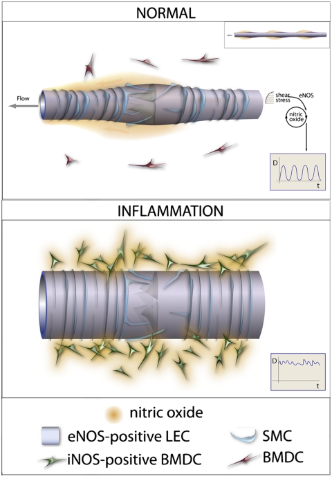
To trigger an effective immune response, antigen and antigen-presenting cells travel to the lymph nodes via collecting lymphatic vessels. However, our understanding of the regulation of collecting lymphatic vessel function and lymph transport is limited. To dissect the molecular control of lymphatic function, we developed a unique mouse model that allows intravital imaging of autonomous lymphatic vessel contraction. Using this method, we demonstrated that endothelial nitric oxide synthase (eNOS) in lymphatic endothelial cells is required for robust lymphatic contractions under physiological conditions. By contrast, under inflammatory conditions, inducible NOS (iNOS)-expressing CD11b(+)Gr-1(+) cells attenuate lymphatic contraction. This inhibition of lymphatic contraction was associated with a reduction in the response to antigen in a model of immune-induced multiple sclerosis. These results suggest the suppression of lymphatic function by the CD11b(+)Gr-1(+) cells as a potential mechanism of self-protection from autoreactive responses during on-going inflammation. The central role for nitric oxide also suggests that other diseases such as cancer and infection may also mediate lymphatic contraction and thus immune response. Our unique method allows the study of lymphatic function and its molecular regulation during inflammation, lymphedema, and lymphatic metastasis.
Conflict of interest statement
Conflict of interest statement: R.K.J. received commercial research grants from Dyax, MedImmune, and Roche; consultant fees from Dyax, SynDevRx, Xtuit, and Noxxon Pharma; and a speaker honorarium from MPM Capital. R.K.J. owns stock in SynDevRx. No reagents or funding from these companies were used in these studies.
Figures
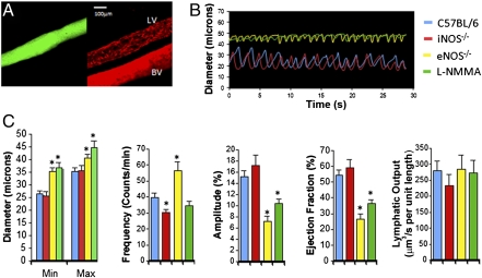
 , C57BL/6, n = 21;
  , iNOS−/−, n = 14;
  , eNOS−/−, n = 27;
  ,
, 
 , C57BL/6, n = 14;
  , iNOS−/−, n = 15;
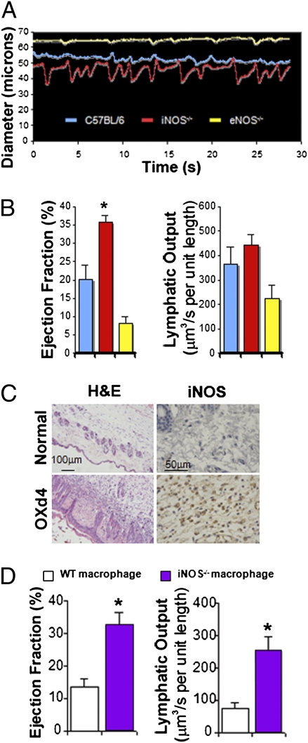
  , eNOS−/−, n = 9. (C) Immunohistochemical staining shows a large cell infiltrate of iNOS+ cells into the s.c. area of inflamed skin (OXd4). (D) Mice implanted with iNOS−/− macrophages maintain strong lymphatic contractions compared with control.
  , WT macrophage, n = 8;
  , iNOS−/− macrophage, n = 8. *P < 0.05. P values are listed in
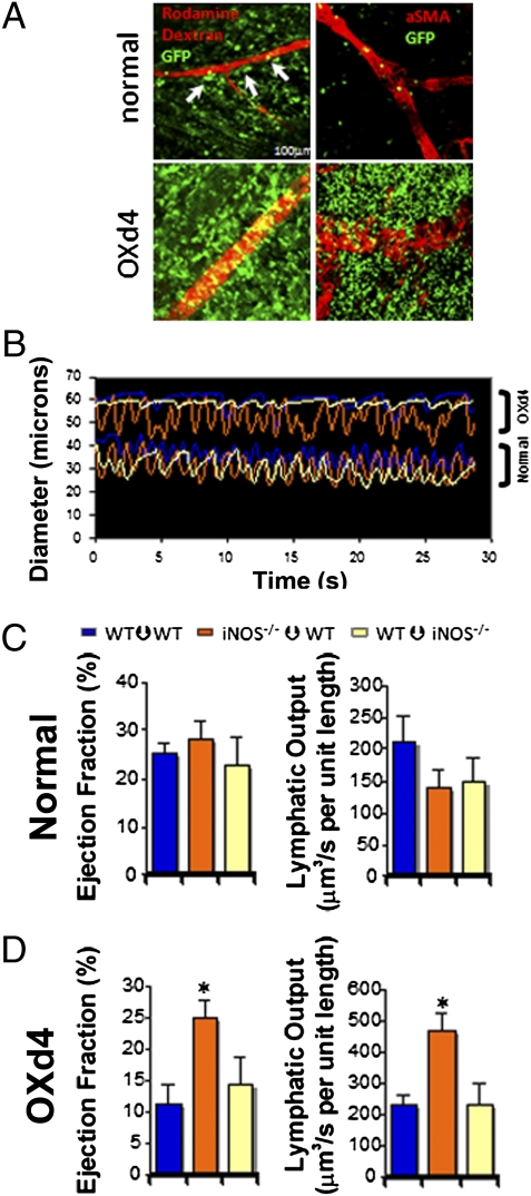
 , WT–WT normal, n = 8, OXd4, n = 7;
  , iNOS−/−–WT normal, n = 8, OXd4, n = 9;
  , WT–iNOS−/− normal, n = 8, OXd4, n = 8. *P < 0.05. P values are listed in
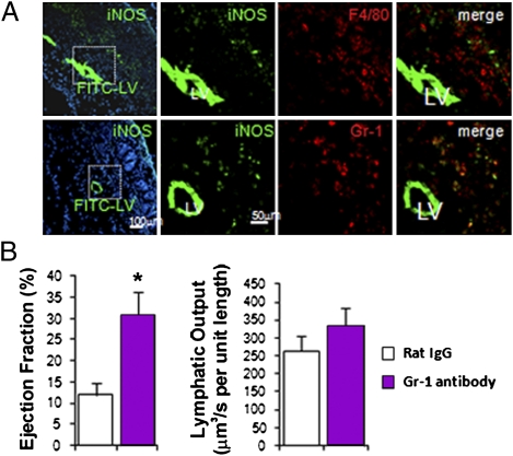
 , Control IgG, n = 8;
  , Gr-1 antibody, n = 9. *P < 0.05.
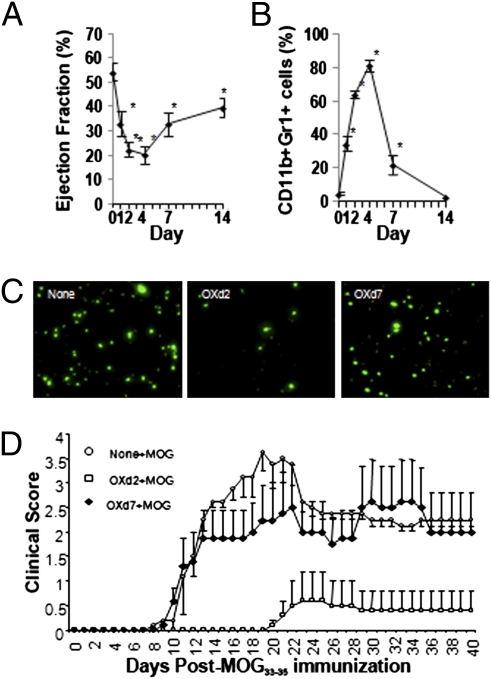
Comment in
-
Nitric oxide (NO) side of lymphatic flow and immune surveillance.Proc Natl Acad Sci U S A. 2012 Jan 3;109(1):3-4. doi: 10.1073/pnas.1117710109. Epub 2011 Dec 16. Proc Natl Acad Sci U S A. 2012. PMID: 22178757 Free PMC article. No abstract available.
References
-
- Jeon BH, et al. Profound but dysfunctional lymphangiogenesis via vascular endothelial growth factor ligands from CD11b+ macrophages in advanced ovarian cancer. Cancer Res. 2008;68:1100–1109. - PubMed
-
- Liao S, Ruddle NH. Synchrony of high endothelial venules and lymphatic vessels revealed by immunization. J Immunol. 2006;177:3369–3379. - PubMed
-
- Hoshida T, et al. Imaging steps of lymphatic metastasis reveals that vascular endothelial growth factor-C increases metastasis by increasing delivery of cancer cells to lymph nodes: Therapeutic implications. Cancer Res. 2006;66:8065–8075. - PubMed
-
- Angeli V, et al. B cell-driven lymphangiogenesis in inflamed lymph nodes enhances dendritic cell mobilization. Immunity. 2006;24:203–215. - PubMed
-
- Padera TP, et al. Lymphatic metastasis in the absence of functional intratumor lymphatics. Science. 2002;296:1883–1886. - PubMed
Publication types
MeSH terms
Substances
Grants and funding
- R01-CA96915/CA/NCI NIH HHS/United States
- R01 CA126642/CA/NCI NIH HHS/United States
- U01 CA084301/CA/NCI NIH HHS/United States
- R00 CA137167/CA/NCI NIH HHS/United States
- R01 CA085140/CA/NCI NIH HHS/United States
- R01-CA115767/CA/NCI NIH HHS/United States
- P01-CA80124/CA/NCI NIH HHS/United States
- K99-CA137167/CA/NCI NIH HHS/United States
- R01 DK057731/DK/NIDDK NIH HHS/United States
- U01-CA084301/CA/NCI NIH HHS/United States
- R01-CA85140/CA/NCI NIH HHS/United States
- R01 CA115767/CA/NCI NIH HHS/United States
- P01 CA080124/CA/NCI NIH HHS/United States
- R01-CA126642/CA/NCI NIH HHS/United States
- R01 CA096915/CA/NCI NIH HHS/United States
- R01 DK57731/DK/NIDDK NIH HHS/United States
- K99 CA137167/CA/NCI NIH HHS/United States
LinkOut - more resources
Full Text Sources
Other Literature Sources
Molecular Biology Databases
Research Materials
