. 2011 Dec 6;108(49):E1321-9.
doi: 10.1073/pnas.1109926108. Epub 2011 Nov 7.

Role of type 2 deiodinase in response to acute lung injury (ALI) in mice

Affiliations

Role of type 2 deiodinase in response to acute lung injury (ALI) in mice

Olga Barca-Mayo et al. Proc Natl Acad Sci U S A. .

Abstract

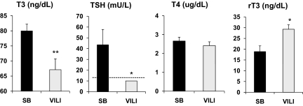
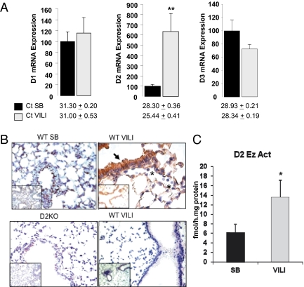
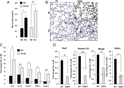
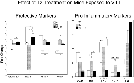
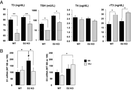
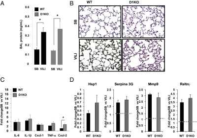
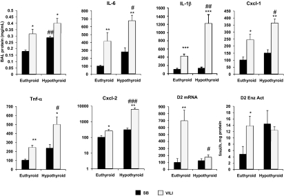
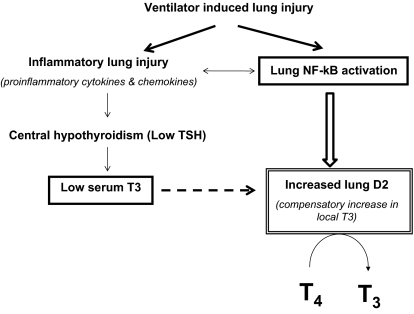
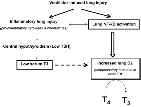
Thyroid hormone (TH) metabolism, mediated by deiodinase types 1, 2, and 3 (D1, D2, and D3) is profoundly affected by acute illness. We examined the role of TH metabolism during ventilator-induced lung injury (VILI) in mice. Mice exposed to VILI recapitulated the serum TH findings of acute illness, namely a decrease in 3,5,3'-triiodothyronine (T(3)) and thyroid-stimulating hormone and an increase in reverse T(3). Both D2 immunoreactivity and D2 enzymatic activity were increased significantly. D1 and D3 activity did not change. Using D2 knockout (D2KO) mice, we determined whether the increase in D2 was an adaptive response. Although similar changes in serum TH levels were observed in D2KO and WT mice, D2KO mice exhibited greater susceptibility to VILI than WT mice, as evidenced by poorer alveoli integrity and quantified by lung chemokine and cytokine mRNA induction. These data suggest that an increase in lung D2 is protective against VILI. Similar findings of increased inflammatory markers were found in hypothyroid WT mice exposed to VILI compared with euthyroid mice, indicating that the lungs were functionally hypothyroid. Treatment of D2KO mice with T(3) reversed many of the lung chemokine and cytokine profiles seen in response to VILI, demonstrating a role for T(3) in the treatment of lung injury. We conclude that TH metabolism in the lung is linked to the response to inflammatory injury and speculate that D2 exerts its protective effect by making more TH available to the injured lung tissue.


Conflict of interest statement

The authors declare no conflict of interest.

Figures

Fig. 1.
Fig. 1.
Serum TH and TSH levels in WT mice after VILI. Serum T3, TSH, T4, and rT3 concentrations in WT mice exposed to VILI (gray bars) compared with SB animals (black bars). Note that TSH levels were undetectable after VILI and were assigned a value equal to the sensitivity of the assay (10 mU/L, indicated by a dashed line) for statistical analysis. Values shown are mean ± SEM. Differences between groups are indicated: *P < 0.05; **P < 0.01. n = 8 animals per group.
Fig. 2.
Fig. 2.
D1, D2, and D3 mRNA levels and D2 protein levels and enzymatic activity in the lungs of WT animals exposed to VILI. (A) Lung D1, D2, and D3 mRNA levels in WT mice after VILI (gray bars) compared with SB animals (black bars). Values shown are mean ± SEM. Significant differences between groups are indicated: *P < 0.05; **P < 0.01. n = 8 animals per group. The number of cycles in the quantitative PCR (Ct) is given below the bars to indicate the relative abundance of each enzyme. (B) (Upper Left) Immunohistochemical staining of D2 (brown) is shown for an SB WT mouse in which D2 protein was graded as 1+ in the epithelium and 1+ in pulmonary endothelium. Minimal inflammatory cells were observed. (Upper Right) Sections from a WT mouse exposed to VILI demonstrate 3+ D2 in the epithelium and 3+ in pulmonary endothelium. More inflammatory cells were observed, but no immunoreactivity was observed in these cells (indicated by asterisk). (Lower Left) Staining of D2 in a section from a D2KO mouse. The absence of D2 staining shows the specificity of the antibody. (Lower Right) Negligible D2 staining of a WT mice after VILI by using the preimmune serum (n = 2 animals per group). Inset shows a lower magnification a representative area of lung. (C) D2 enzymatic activity after VILI (gray bar) compared with SB animals (black bar). Values shown are mean ± SEM. Significant differences between groups are indicated: *P < 0.05. n = 8 animals per group.
Fig. 3.
Fig. 3.
D2KO mice have increased susceptibility to VILI. (A) BAL protein content in D2KO mice compared with WT mice exposed to VILI. VILI evoked significant elevations in BAL protein levels in WT (*P < 0.05) and D2KO mice (**P < 0.01). (B) H&E staining of representative lung sections of SB and VILI-exposed WT and D2KO mice. No pathological differences were seen between SB WT and D2KO mice. Cell infiltration in the alveolar wall and hyaline membrane formation were increased in WT mice exposed to VILI (arrows). Histology of a representative D2KO mouse exposed to VILI that shows higher polymorphonuclear cells infiltration and hyaline membrane formation compared with their correspondent WT mice (three animals per group). (C) Fold change of cytokines and chemokines (IL-6, IL-1β, Cxcl1, TNF-α and Cxcl2) by qPCR in WT mice (n = 6) (black bars) or D2KO mice (n = 6) (gray bars) exposed to VILI relative to SB animals as control. Differences between groups are indicated: *P < 0.05; **P < 0.01. (D) Fold change of Hsp1, Serpina 3G, Mmp9, and Retlnγ by qPCR in WT mice (n = 8) (black bars) or D2KO mice (n = 6) (gray bars) exposed to VILI relative to SB animals as control. Differences between groups are indicated: *P < 0.05; **P < 0.01. Dashed line is drawn at a fold change of “1” for purposes of comparison.
Fig. 4.
Fig. 4.
Effect of l-T3 treatment on cytokine and chemokine expression in lungs of D2KO mice after VILI. Mice treated with 0.2 μg/100 g l-T3 for 5 d were subjected to VILI or SB 2 h after the last injection. Shown is the fold change in markers of inflammation and injury by qPCR in WT (n = 6), D2KO (n = 6), and l-T3–treated D2KO (n = 6) mice exposed to VILI relative to SB animals as control. In general, protective markers of inflammation (Serpina 3G, Hsp1) increased with l-T3 treatment (although Mmp9 and Retnlγ did not), and proinflammatory markers (Cxcl1, TNF, IL-1β, and Cxcl2) were suppressed with l-T3 (although IL-6 was not). Differences between groups are indicated: *P < 0.05; **P < 0.01.
Fig. 5.
Fig. 5.
Serum TH and TSH concentrations and D1 and D3 mRNA expression in D2KO mice after VILI. (A) Serum T3, TSH, T4, and rT3 concentrations in ventilated WT and D2KO mice (gray bars) compared with SB animals (black bars). Note that after VILI the TSH levels in WT mice were undetectable and were assigned a value equal to the sensitivity of the assay (10 mU/L, indicated by a dashed line) for statistical analysis. Values shown are mean ± SEM. *P < 0.05; **P < 0.01 VILI vs. SB. n = 6 animals per group. (B) Lung D1 and D3 mRNA levels in WT and D2KO mice after VILI compared with SB animals as control. n = 8 animals per group. Values shown are ± SEM. Significant differences between groups are indicated: *P < 0.05; #P < 0.05 vs. WT SB.
Fig. 6.
Fig. 6.
Response to VILI is comparable in D1KO and WT mice. (A) BAL protein content in WT and D1KO mice exposed to VILI compared with SB animals. Differences between groups are indicated. *P < 0.05. (B) H&E staining of a lung section of a WT mouse and a D1KO mouse SB and after exposure to VILI. Histology of VILI-exposed WT and D1KO mice shows higher polymorphonuclear cell infiltration and hyaline membrane formation with no differences between the two groups (n = 3 animals per group). (C) Fold changes of VILI biomarker genes (IL-6, IL-1β, Cxcl1, TNF-α, and Cxcl2) by qPCR in WT and D1KO mice exposed to VILI relative to SB animals as control. *P < 0.05 vs. WT mice. (D) Fold change of Hsp1, Serpina 3G, Mmp9, and Retlnγ by qPCR in WT (n = 8) and D1KO (n = 6) (gray bars) mice exposed to VILI relative to SB animals as control.
Fig. 7.
Fig. 7.
Hypothyroid WT mice have increased susceptibility to VILI. BAL protein content in hypothyroid mice compared with euthyroid animals exposed to VILI. VILI evoked significant elevations in BAL protein levels in euthyroid mice (*P < 0.05) and hypothyroid mice (*P < 0.05; ##P < 0.01 vs. SB euthyroid mice). mRNA levels of the VILI biomarker genes IL-6, IL-1β, Cxcl1, TNF-α, and Cxcl by qPCR in euthyroid (n = 8) and hypothyroid (n = 6) mice SB and after VILI. Lung D2 mRNA and enzymatic activity (Enz Act) in euthyroid and hypothyroid mice after VILI compared with SB animals as control. n = 6 animals per group. Values shown are mean ± SEM. *P < 0.05 vs. SB; **P < 0.01 vs. SB; ***P < 0.001 vs. SB; #P < 0.05 vs. euthyroid VILI; ##P < 0.01 vs. euthyroid VILI; ###P < 0.001 vs. euthyroid VILI.
Fig. 8.
Fig. 8.
Model of lung D2 induction by VILI. VILI is characterized by increases in inflammatory cytokine expression and activation of mediators of immune and inflammatory responses such as NF-κB. The proinflammatory cytokines increase hypothalamic D2 (17) and cause central hypothyroidism that might be responsible for the low serum T3. The induction of D2 in the lung by mechanical ventilation might be adaptive to the low circulating levels of T3 or in response to the local activation of NF-κB. The dashed arrow represents the posttranslational effect of T3 on D2.
Fig. P1.
Fig. P1.
Ventilator-induced lung injury is characterized by increases in inflammatory cytokine expression and activation of mediators of immune and inflammatory responses such as NF-κB. Proinflammatory cytokines increase hypothalamic D2 and cause central hypothyroidism, which is likely responsible for the low serum levels of T3. The induction of D2 in the lung by mechanical ventilation appears to be an adaptation to the low circulating levels of T3 or may caused by to the local activation of NF-κB. The dashed arrow represents the posttranslational effect of T3 on D2.

References

    1. Bianco AC, Salvatore D, Gereben B, Berry MJ, Larsen PR. Biochemistry, cellular and molecular biology, and physiological roles of the iodothyronine selenodeiodinases. Endocr Rev. 2002;23(1):38–89. - PubMed
    1. Alkemade A. Central and peripheral effects of thyroid hormone signalling in the control of energy metabolism. J Neuroendocrinol. 2010;22(1):56–63. - PubMed
    1. Williams GR. Neurodevelopmental and neurophysiological actions of thyroid hormone. J Neuroendocrinol. 2008;20:784–794. - PubMed
    1. Gogakos AI, Duncan Bassett JH, Williams GR. Thyroid and bone. Arch Biochem Biophys. 2010;503(1):129–136. - PubMed
    1. Ness GC. Thyroid hormone. Basis for its hypocholesterolemic effect. J Fla Med Assoc. 1991;78:383–385. - PubMed

Publication types

MeSH terms

LinkOut - more resources