Comparative Study
. 2011 Nov 29;108(48):E1244-53.
doi: 10.1073/pnas.1111255108. Epub 2011 Nov 7.

Lower specific infectivity of protease-resistant prion protein generated in cell-free reactions

Affiliations
Comparative Study

Lower specific infectivity of protease-resistant prion protein generated in cell-free reactions

Mikael Klingeborn et al. Proc Natl Acad Sci U S A. .

Abstract

Prions are unconventional infectious agents that cause transmissible spongiform encephalopathy (TSE) diseases, or prion diseases. The biochemical nature of the prion infectious agent remains unclear. Previously, using a protein misfolding cyclic amplification (PMCA) reaction, infectivity and disease-associated protease-resistant prion protein (PrPres) were both generated under cell-free conditions, which supported a nonviral hypothesis for the agent. However, these studies lacked comparative quantitation of both infectivity titers and PrPres, which is important both for biological comparison with in vivo-derived infectivity and for excluding contamination to explain the results. Here during four to eight rounds of PMCA, end-point dilution titrations detected a >320-fold increase in infectivity versus that in controls. These results provide strong support for the hypothesis that the agent of prion infectivity is not a virus. PMCA-generated samples caused the same clinical disease and neuropathology with the same rapid incubation period as the input brain-derived scrapie samples, providing no evidence for generation of a new strain in PMCA. However, the ratio of the infectivity titer to the amount of PrPres (specific infectivity) was much lower in PMCA versus brain-derived samples, suggesting the possibility that a substantial portion of PrPres generated in PMCA might be noninfectious.


Conflict of interest statement

The authors declare no conflict of interest.

Figures

Fig. 1.
Fig. 1.
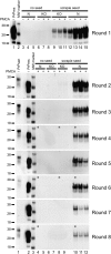
Amplification of tg7 hamster PrPres by eight rounds of serial PMCA. (A and B) tg7 experiment 1 (A) and tg7 experiment 2 (B). A 10% brain homogenate from a 263K scrapie-infected tg7 mouse was diluted 10-fold, i.e., 1:100 final brain dilution, into 10% normal brain homogenate from perfused healthy tg7 mice. Following a first round [round 1 (R1)] of PMCA, the sample was diluted 10-fold (5 μL into 45 μL) into 10% tg7 normal brain homogenate for a second round (R2) of PMCA, and this was repeated for eight rounds. After each round 5 μL of each 50-μL PMCA reaction tube was PK digested and analyzed by Western blotting. The PrP substrate in the normal brain homogenate is shown by a non-PK–treated sample, indicated as PrPsen in lane 1 (0.05 mg tissue equivalent), and the seed brain used in these serial PMCA experiments is indicated as PrPres in lane 2 (0.25 mg tissue equivalent). Unseeded reactions of normal tg7 brain homogenate (N), unseeded reactions of PrP knockout mouse brain homogenate (KO), seeded reactions with KO substrate, and seeded reactions with N substrate are indicated. Compared with freeze-control samples that were not subjected to PMCA sonication and incubation (indicated with a “−” sign), products from R1 showed an average amplification of 32- and 12-fold in round 1 of tg7 experiment (expt.) 1 (A) and tg7 expt. 2 (B), respectively. Similar unsonicated freeze controls were done for each round and indicated amplification ranging from 4- to 32-fold in rounds 2–8. A PrPres signal higher than detected in the freeze-control samples is indicative of newly generated PrPres [in R1: (A) compare lane 8 with lanes 9–11 and (B) compare lane 12 with lanes 13–15]. Samples from rounds 7 and 8 in Fig. 1A were run on the same gel and to aid presentation, controls in lanes 1–3 are shown in both round 7 and round 8. Samples used for animal infectivity bioassays are indicated by asterisks. Apparent molecular masses of 18, 28, and 38 kDa are indicated on the left.
Fig. 2.
Fig. 2.
End-point titration of brain-derived and PMCA-generated prion infectivity in tg7 mice. Incubation time is shown on the y axis as days postinoculation (dpi) and 10-fold dilutions of the inoculum are shown on the x axis. Dilutions are shown relative to samples in the PMCA reaction tubes. (A–G) Infectivity data from tg7 mice inoculated with (A) brain-derived tg7 263K scrapie diluted 1:100 in the R1 freeze-control tube as described or (B–G) PMCA products from two separate tg7 PMCA experiments. PMCA products from round 4 (B and E), round 6 (C and F), and round 8 (D and G) were tested. Solid symbols (•) represent animals that died or showed clinical neurological signs. Scrapie was confirmed by detection of PrPres in the brain by Western blot. Open symbols (○) represent animals that did not contract disease. To aid presentation, animals that succumbed to intercurrent death and tested negative for PrPres are not shown.
Fig. 3.
Fig. 3.
Amplification of Syrian hamster (SHa) PrPres by serial PMCA. A 10% brain homogenate from a 263K scrapie-infected SHa was diluted 100-fold, i.e., 1:1,000 final brain dilution, into 10% normal brain homogenate from perfused healthy SHa. Following a first round of PMCA, the sample was diluted 10-fold into 10% SHa normal brain homogenate for a second round of PMCA. This process of serial PMCA was repeated for a total of eight rounds. A total of 5 μL of each 50-μL PMCA reaction tube was PK digested and then analyzed by Western blotting. The PrP substrate in the normal brain homogenate is shown by a non-PK–treated sample, indicated as PrPsen in lane 1 in rounds 2–8 (0.05 mg tissue equivalent). The seed used in this serial PMCA is indicated as PrPres in lane 1 in round 1 and lane 2 in rounds 2–8 (0.05 mg tissue equivalent). Duplicate unseeded reactions of normal hamster brain homogenate (N) and PrP KO mouse brain homogenate and seeded reactions with KO in duplicate and N in triplicate are indicated. Compared with freeze-control samples that were not subjected to PMCA (indicated with a “−” sign), products from R1 showed a 6-fold amplification. Similar freeze controls were done for each round and indicated an amplification ranging from 3- to 16-fold in rounds 2–8. A PrPres signal higher than detected in the freeze-control samples is indicative of newly generated PrPres (compare lane 12 with lanes 13–15 in R1). Samples used for animal infectivity bioassays are indicated by asterisks. Apparent molecular masses of 18, 28, and 38 kDa are indicated on the left.
Fig. 4.
Fig. 4.
End-point titration of brain-derived and PMCA-generated prion infectivity in hamsters. Incubation time is shown on the y axis as days postinoculation (dpi) and 10-fold dilutions of the inoculum are shown on the x axis. Dilutions are shown relative to samples in the PMCA reaction tubes. Syrian hamsters were inoculated with brain-derived 263K scrapie (A) and PMCA products from round 4 (B), round 6 (C), and round 8 (D). Solid symbols (•) represent animals that died or showed clinical neurological signs. Scrapie was confirmed by detection of PrPres in the brain by Western blot. Open symbols (○) represent animals that did not contract disease. To aid presentation, animals that succumbed to intercurrent death and tested negative for PrPres are not shown.
Fig. 5.
Fig. 5.
Comparison of specific infectivity (infectivity in LD50/PrPres units) in brain-derived (R1 input) vs. PMCA-derived (output from R4, R6, and R8) materials. Results from two experiments with tg7 brain and one experiment with hamster brain are shown. Infectivity was quantitated in rounds 4, 6, and 8 of the PMCA reaction by in vivo endpoint dilution titration. In all experiments new infectivity was generated during PMCA reactions; however, the specific infectivity was much lower for PMCA-derived material than for brain-derived material. The SEs for the specific infectivity values were calculated as described in Materials and Methods. Each experiment was analyzed statistically by one-way ANOVA with Tukey's multiple comparison test, using GraphPad Prism software. P values shown are for comparison of R1 input brain-derived material vs. PMCA-derived material from each round tested: ***P < 0.001; **P < 0.01; *P < 0.05.
Fig. 6.
Fig. 6.
Spongiosis and PrPres deposition in brains of animals inoculated with brain-derived or PMCA-generated prion infectivity. Tg7 hamster PrP transgenic mice (A–D) and Syrian hamsters (E and F) inoculated with brain-derived and PMCA-generated prion infectivity were scored for severity of spongiform change (A, C, and E) and amount of PrPres deposition (B, D, and F) in 10 indicated brain areas. Tg7 experiment 1 is shown in A and B, and experiment 2 in C and D. The number of animals scored in each group is indicated in parentheses in the Inset and the scoring procedures are described in Materials and Methods. There was overlap of SD between groups, indicating that there were no statistically significant differences. Therefore, SD bars were omitted as they interfered with the presentation.
Fig. P1.
Fig. P1.
Comparison of specific infectivity (infectivity LD50/PrPres units) in brain-derived vs. PMCA-derived materials. Results represent data from one experiment using brains of tg7 mice genetically engineered to express prion protein encoded by hamster genes. Similar results were obtained in a second experiment using tg7 brain and in a third experiment using hamster brain. Infectivity concentration was quantified for the brain-derived input and the PMCA products obtained after specific numbers of rounds of PMCA by standard end-point dilution measurement methods using tg7 mice. In all experiments, new infectivity was generated during PMCA reactions; however, the ratio of infectivity to PrPres (specific infectivity) was much lower for PMCA-derived material than for brain-derived material.

Comment in

  • Viruses do replicate in cell-free systems.
    Karapetyan YE. Karapetyan YE. Proc Natl Acad Sci U S A. 2012 Feb 21;109(8):E461; author reply E462. doi: 10.1073/pnas.1118908109. Epub 2012 Jan 30. Proc Natl Acad Sci U S A. 2012. PMID: 22308429 Free PMC article. No abstract available.

Similar articles

Cited by

References

    1. Chesebro B. Introduction to the transmissible spongiform encephalopathies or prion diseases. Br Med Bull. 2003;66:1–20. - PubMed
    1. Brandner S, et al. Normal host prion protein (PrPC) is required for scrapie spread within the central nervous system. Proc Natl Acad Sci USA. 1996;93:13148–13151. - PMC - PubMed
    1. Büeler H, et al. Mice devoid of PrP are resistant to scrapie. Cell. 1993;73:1339–1347. - PubMed
    1. Rohwer RG. The scrapie agent: “a virus by any other name”. In: Chesebro BW, editor. Transmissible Spongiform Encephalopathies, Scrapie, BSE and Related Disorders, Current Topics in Microbiology and Immunology. Vol. 172. New York: Springer; 1991. pp. 195–232. - PubMed
    1. Griffith JS. Self-replication and scrapie. Nature. 1967;215:1043–1044. - PubMed

Publication types

LinkOut - more resources