Nanoparticle-mediated signaling endosome localization regulates growth cone motility and neurite growth
- PMID: 22065745
- PMCID: PMC3223462
- DOI: 10.1073/pnas.1019624108
Nanoparticle-mediated signaling endosome localization regulates growth cone motility and neurite growth
Abstract
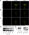
Understanding neurite growth regulation remains a seminal problem in neurobiology. During development and regeneration, neurite growth is modulated by neurotrophin-activated signaling endosomes that transmit regulatory signals between soma and growth cones. After injury, delivering neurotrophic therapeutics to injured neurons is limited by our understanding of how signaling endosome localization in the growth cone affects neurite growth. Nanobiotechnology is providing new tools to answer previously inaccessible questions. Here, we show superparamagnetic nanoparticles (MNPs) functionalized with TrkB agonist antibodies are endocytosed into signaling endosomes by primary neurons that activate TrkB-dependent signaling, gene expression and promote neurite growth. These MNP signaling endosomes are trafficked into nascent and existing neurites and transported between somas and growth cones in vitro and in vivo. Manipulating MNP-signaling endosomes by a focal magnetic field alters growth cone motility and halts neurite growth in both peripheral and central nervous system neurons, demonstrating signaling endosome localization in the growth cone regulates motility and neurite growth. These data suggest functionalized MNPs may be used as a platform to study subcellular organelle localization and to deliver nanotherapeutics to treat injury or disease in the central nervous system.
Conflict of interest statement
The authors declare no conflict of interest.
Figures


Similar articles
-
Signaling endosomes and growth cone motility in axon regeneration.Int Rev Neurobiol. 2012;106:35-73. doi: 10.1016/B978-0-12-407178-0.00003-X. Int Rev Neurobiol. 2012. PMID: 23211459 Review.
-
Rab22 controls NGF signaling and neurite outgrowth in PC12 cells.Mol Biol Cell. 2011 Oct;22(20):3853-60. doi: 10.1091/mbc.E11-03-0277. Epub 2011 Aug 17. Mol Biol Cell. 2011. PMID: 21849477 Free PMC article.
-
Cdc42 stimulates neurite outgrowth and formation of growth cone filopodia and lamellipodia.J Neurobiol. 2000 Jun 15;43(4):352-64. doi: 10.1002/1097-4695(20000615)43:4<352::aid-neu4>3.0.co;2-t. J Neurobiol. 2000. PMID: 10861561
-
Mitochondrial dynamics regulate growth cone motility, guidance, and neurite growth rate in perinatal retinal ganglion cells in vitro.Invest Ophthalmol Vis Sci. 2012 Oct 30;53(11):7402-11. doi: 10.1167/iovs.12-10298. Invest Ophthalmol Vis Sci. 2012. PMID: 23049086 Free PMC article.
-
Micro-scale chromophore-assisted laser inactivation of nerve growth cone proteins.Microsc Res Tech. 2000 Jan 15;48(2):97-106. doi: 10.1002/(SICI)1097-0029(20000115)48:2<97::AID-JEMT5>3.0.CO;2-G. Microsc Res Tech. 2000. PMID: 10649510 Review.
Cited by
-
Biophysical Tools for Cellular and Subcellular Mechanical Actuation of Cell Signaling.Biophys J. 2016 Sep 20;111(6):1112-1118. doi: 10.1016/j.bpj.2016.02.043. Epub 2016 Jul 25. Biophys J. 2016. PMID: 27456131 Free PMC article. Review.
-
Manipulation of New Fluorescent Magnetic Nanoparticles with an Electromagnetic Needle, Allowed Determining the Viscosity of the Cytoplasm of M-HeLa Cells.Pharmaceuticals (Basel). 2023 Jan 29;16(2):200. doi: 10.3390/ph16020200. Pharmaceuticals (Basel). 2023. PMID: 37259349 Free PMC article.
-
Nanoparticle mediated drug delivery of rolipram to tyrosine kinase B positive cells in the inner ear with targeting peptides and agonistic antibodies.Front Aging Neurosci. 2015 May 19;7:71. doi: 10.3389/fnagi.2015.00071. eCollection 2015. Front Aging Neurosci. 2015. PMID: 26042029 Free PMC article.
-
Electromagnetic Regulation of Cell Activity.Cold Spring Harb Perspect Med. 2019 May 1;9(5):a034322. doi: 10.1101/cshperspect.a034322. Cold Spring Harb Perspect Med. 2019. PMID: 30249601 Free PMC article. Review.
-
An atlas of nano-enabled neural interfaces.Nat Nanotechnol. 2019 Jul;14(7):645-657. doi: 10.1038/s41565-019-0487-x. Epub 2019 Jul 3. Nat Nanotechnol. 2019. PMID: 31270446 Free PMC article. Review.
References
-
- Goldberg JL, Klassen MP, Hua Y, Barres BA. Amacrine-signaled loss of intrinsic axon growth ability by retinal ganglion cells. Science. 2002;296:1860–1864. - PubMed
-
- Nash M, Pribiag H, Fournier AE, Jacobson C. Central nervous system regeneration inhibitors and their intracellular substrates. Mol Neurobiol. 2009;40:224–235. - PubMed
Publication types
MeSH terms
Substances
Grants and funding
LinkOut - more resources
Full Text Sources
Other Literature Sources

