. 2011 Nov 15;108(46):18672-7.
doi: 10.1073/pnas.1110415108. Epub 2011 Nov 7.

Insights regarding guanine nucleotide exchange from the structure of a DENN-domain protein complexed with its Rab GTPase substrate

Affiliations

Insights regarding guanine nucleotide exchange from the structure of a DENN-domain protein complexed with its Rab GTPase substrate

Xudong Wu et al. Proc Natl Acad Sci U S A. .

Abstract

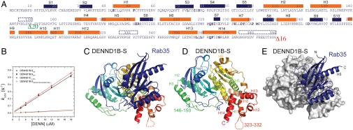
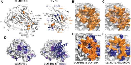
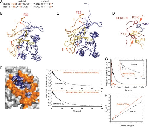
Rab GTPases are key regulators of membrane traffic pathways within eukaryotic cells. They are specifically activated by guanine nucleotide exchange factors (GEFs), which convert them from their "inactive" GDP-bound form to the "active" GTP-bound form. In higher eukaryotes, proteins containing DENN-domains comprise a major GEF family. Here we describe at 2.1-Å resolution the first structure of a DENN-domain protein, DENND1B-S, complexed with its substrate Rab35, providing novel insights as to how DENN-domain GEFs interact with and activate Rabs. DENND1B-S is bi-lobed, and interactions with Rab35 are through conserved surfaces in both lobes. Rab35 binds via switch regions I and II, around the nucleotide-binding pocket. Positional shifts in Rab residues required for nucleotide binding may lower its affinity for bound GDP, and a conformational change in switch I, which makes the nucleotide-binding pocket more solvent accessible, likely also facilitates exchange.


Conflict of interest statement

The authors declare no conflict of interest.

Figures

Fig. 1.
Fig. 1.
Structure of the DENND1B-S/Rab35 complex. (A) Sequence of DENND1B-S in crystallization construct. Secondary structure as in the crystal structure is indicated. Helices are orange bars and strands blue. Disordered regions are indicated by dotted lines. Sixteen residues at the C terminus and a 20-residue loop in wild-type protein were deleted; their locations are indicated by green and red arrowheads. (B) The DENND1B-S construct used for crystallization is equally efficient in catalyzing guanine nucleotide exchange for Rab35 as wild-type protein. MantGDP dissociation from Rab35 was continuously monitored by FRET (λex = 280 nm, λem = 435 nm) with increasing concentrations of either the crystallization construct or wild-type DENN domain. The DENND1C DENN domain is approximately 10-fold less efficient, as is a DENND1B mutant, where residues at the Rab35 interface were changed to corresponding residues in DENND1C. See Fig. S5A for representative time course data. (C) Ribbons diagram of DENND1B-S, colored with blue at the N terminus and red at the C terminus, complexed with Rab35 (blue). (D) DENND1B-S alone with secondary structure elements labeled. (E) Rab35 is rendered as a ribbon, DENND1B-S as a surface representation.
Fig. 2.
Fig. 2.
Interactions between DENND1B-S and Rab35. (A) Residues in DENND1B-S within 5 Å of Rab35 are labeled (left), as are residues in Rab35 within 5 Å of the DENN-domain (right). In Rab35, interacting residues in switches I or II are orange and residues outside the switch region blue. A 180° rotation about a vertical axis would position the Rab to interact with DENND1B-S. (B) DENND1B-S oriented as in A with residues within 4.5 Å of Rab35 in orange and labeled. (C) DENND1B-S as in B, with Rab35 switch regions I and II bound. Rab35 residues are labeled. (D) Sequence conservation in 18 human DENN domains mapped onto the surface of DENND1B-S. Strictly, highly, and weakly conserved residues (identical in 16–18, 11–15, and 8–10 sequences, respectively) are purple, magenta, and lilac, respectively. (E) The Rab35 interaction surface is similar in DENND1B and −1A. The Rab35 interaction surface on DENND1B is as in B, with residues in DENND1B within 10 Å of Rab35 that are different in DENND1A indicated (blue). Residues further than 5 Å from Rab35 are in parentheses. (F) As in E but comparing DENND1B and −1C.
Fig. 3.
Fig. 3.
Rearrangement of Rab35 and switches I and II in the Rab/GEF complex. (A) Sequences in the switch regions of Rab35 and Rab1 are highly conserved. See also Fig. S2F for a comparison of Rab1 and Rab35 surfaces. (B) Comparison of Rab35 (purple) in the complex with GDP-bound Rab1 (lilac; PDB ID code 2FOL ). Switches I and II are orange or yellow, respectively, and GDP and Mg2+ are yellow. F33 (Y33 in Rab1) moves out of the nucleotide-binding pocket as switch I alters its conformation. F33 is not well ordered and that the position of its side chain is uncertain. Arrows indicate conformational changes. (C) As in B but comparing Rab35 in the complex with GTP-bound Rab1 (PDB ID code 1YZN). (D) Switch I in Rab35 must rearrange as Rab35 binds DENND1B because it would otherwise clash sterically with H7 in the DENN domain (brown). Rab35 in the Rab/GEF complex is purple/orange; GDP-bound Rab is lilac/yellow. Arrows indicate clashes. (E) DENND1B residues predicted to affect either switch I or II binding were mutated, as indicated (blue and lilac for I and II, respectively). Compare this panel to Fig. 2 BC. (F) The catalytic efficiency of the mutants is reduced compared to wild type as assessed by monitoring mantGDP release. A representative time course with DENN domains at 4 μM. (G) [3H]GDP dissociates approximately 20-fold faster from the Rab35 F33A mutant than from Rab35. A time course of [3H]GDP dissociating from Rab35 or Rab35-F33A (both at 200 nM concentration) is shown. The solid line is the best fit of a single exponential function, yielding a GDP dissociation rate constant (k-GDP) of 1.35 ± 0.07 × 10-4 s-1 for Rab35 and 25.6 ± 2.8 × 10-4 s-1 for Rab35-F33A. (H) mantGDP association rates are similar for Rab35 and the F33A mutant. MantGDP binding to nucleotide-free Rab was monitored by FRET (Fig. S5B). The concentration dependence of the observed GDP association rate constant (k+GDP) on [mantGDP] is shown. The rate constants from the slope of the best linear fit are 2.09 ± 0.03 μM-1 s-1 for Rab35 and 2.59 ± 0.09 μM-1 s-1 for Rab35 (F33A). Data were generated considering only the fast kobs from the double-exponential fit. The true concentration dependence may be hyperbolic, but we were not able to work at sufficiently high mantGDP concentration to see saturation.

References

    1. Cai H, Reinisch K, Ferro-Novick S. Coats, tethers, Rabs, and SNAREs work together to mediate the intracellular destination of a transport vesicle. Dev Cell. 2007;12:671–682. - PubMed
    1. Behnia R, Munro S. Organelle identity and the signposts for membrane traffic. Nature. 2005;438:597–604. - PubMed
    1. Pfeffer SR, Aivazian D. Targeting Rab GTPases to distinct membrane compartments. Nat Rev Mol Cell Biol. 2004;5:886–896. - PubMed
    1. Zerial M, McBride H. Rab proteins as membrane organizers. 2001;2:107–117. - PubMed
    1. Goody RS, Hofmann-Goody W. Exchange factors, effectors, GAPs and motor proteins: Common thermodynamic and kinetic principles for different functions. Eur Biophys J. 2002;31:268–274. - PubMed

Publication types

MeSH terms

Associated data