. 2012 Jan 10;109(2):E68-75.
doi: 10.1073/pnas.1107017108. Epub 2011 Nov 7.

Wip1 promotes RUNX2-dependent apoptosis in p53-negative tumors and protects normal tissues during treatment with anticancer agents

Affiliations

Wip1 promotes RUNX2-dependent apoptosis in p53-negative tumors and protects normal tissues during treatment with anticancer agents

Anastasia R Goloudina et al. Proc Natl Acad Sci U S A. .

Abstract

The inactivation of the p53 tumor suppressor pathway in many cancers often increases their resistance to anticancer therapy. Here we show that a previously proposed strategy directed to Wip1 inhibition could be ineffective in tumors lacking p53. On the contrary, Wip1 overexpression sensitized these tumors to chemotherapeutic agents. This effect was mediated through interaction between Wip1 and RUNX2 that resulted, in response to anticancer treatment, in RUNX2-dependent transcriptional induction of the proapoptotic Bax protein. The potentiating effects of Wip1 overexpression on chemotherapeutic agents were directed only to tumor cells lacking p53. The overexpression of Wip1 in normal tissues provided protection from cisplatin-induced apoptosis through decreased strength of upstream signaling to p53. Thus, Wip1 phosphatase promotes apoptosis in p53-negative tumors and protects normal tissues during treatment with anticancer agents.


Conflict of interest statement

The authors declare no conflict of interest.

Figures
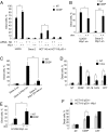
Fig. 1.
Fig. 1.
Wip1 sensitizes tumor cells to chemotherapeutic agents. (A) Down-regulation of Wip1 sensitized cancer cell lines to cisplatin (CDDP) only in the presence of p53. WT p53-expressing cells (human osteosarcoma U2OS cells and colorectal cancer HCT116 cells) and p53-negative cells (human osteosarcoma Saos-2 and colorectal cancer HCT116 p53−/− cells) were transfected with control scrambled siRNA or Wip1 siRNA. Twenty-four hours after transfection, cells were nontreated (NT) or treated with 25 μM CDDP for 48 h, harvested, and subjected to Guava ViaCount cell death assay. (B) Sensitivity of Saos-2 and U2OS cells to cisplatin (CDDP) with or without doxycycline-induced Wip1 expression. Wip1 was induced by doxycycline for 24 h in several clones of established U2OS-Wip1-on and Saos-2-Wip1-on cell lines (Fig. S1B). Cells with or without Wip1 induction were treated with 25 μM cisplatin for 48 h, harvested, and subjected to Guava ViaCount cell death assay (*P < 0.05). (C) Overexpression of phosphatase inactive Wip1 does not sensitize Saos-2 cells to cisplatin. Saos-2 cells were transfected with mutant Wip1 D314A expressing vector DNA; Saos-2 Wip-on cells were cultivated with or without doxycycline. Twenty-four hours later, cells were treated with 25 μM CDDP for 48 h, harvested, and subjected to Guava ViaCount cell death assay. (D) Overexpression of Wip1 sensitizes Saos-2 cells to various anticancer drugs. Saos-2 cells with inducible Wip1 were cultivated with or without doxycycline for 24 h and then treated with 25 μM CDDP, 10 μM etoposide (VP-16), 20 μg/mL of 5-fluorouracil (5-FU), or 1 μM camptothecin (CPT) for 48 h. Cells were harvested and subjected to Guava ViaCount cell death assay. (E) Overexpression of Wip1 sensitizes non–small-cell lung carcinoma H1299 cells to CDDP. H1299 cells with inducible Wip1 were maintained with or without doxycycline for 24 h and then treated with 25 μM CDDP for 48 h. Cells were harvested and subjected to Guava ViaCount cell death assay. (F) Retroviral overexpression of Wip1 sensitizes colon cancer cells HCT116 (p53−/−) to cisplatin. Cells were infected with retrovirus expressing Wip1 and treated with 25 μM CDDP for 48 h, harvested, and subjected to Guava ViaCount cell death assay (*P < 0.05, **P < 0.01, and ***P < 0.001; ns, not statistically significant at the 95% confidence level). Data represent the mean ± SEM of two to four independent experiments.
Fig. 2.
Fig. 2.
Dephosphorylation of activating serines in Chk1 by Wip1 and G2 checkpoint. (A) Wip1 suppresses phosphorylation of Chk1. Saos-2-Wip1-on cells were cultivated with or without doxycycline. Twenty-four hours later, cells were treated with 25 μM cisplatin (CDDP), harvested at indicated time points and analyzed by immunoblotting. Data are representative of three independent experiments. (B) Wip1 overexpression does not abrogate G2 checkpoint. Saos2-Wip1-on cells were cultivated with or without doxycycline. Twenty-four hours later, cells were treated with 25 μM CDDP for 12 h. Nocodazole was added to trap cells in mitosis. Cell were harvested, fixed, and stained for phosphohistone H3 as a mitotic marker. Data represent the mean ± SEM of three independent experiments.
Fig. 3.
Fig. 3.
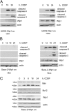
Characterization of cisplatin-induced apoptosis in U2OS-Wip1-on and Saos-2-Wip1-on cells. (A) Induction of Wip1 in U2OS-Wip1-on cells delays caspase 9 and caspase 3 activation after cisplatin (CDDP). U2OS-Wip1-on cells were cultivated with or without doxycycline for 24 h, then treated with 25 μM CDDP for indicated time points, harvested, and analyzed by immunoblotting. (B) Induction of Wip1 in Saos-2-Wip1-on cells leads to strong caspase 9 and caspase 3 activation after CDDP, Saos-2-Wip1-on cells were cultivated with or without doxycycline for 24 h, then treated with 25 μM cisplatin for indicated time points, harvested, and analyzed by immunoblotting. (C) Overexpression of Wip1 results in Bax induction after cisplatin treatment. Saos-2-Wip1-on cells were cultivated with or without doxycycline for 24 h, and then treated with 25 μM cisplatin for indicated time points, harvested, and immunoblotted. Data are representative of three independent experiments.
Fig. 4.
Fig. 4.
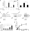
Role of RUNX2 transcriptional factor in Wip1-dependent cisplatin-induced apoptosis. (A) RUNX2 silencing abrogates Wip1-mediated cell death after cisplatin (CDDP) treatment in Saos-2-Wip1-on cells. Saos-2-Wip1-on cells were transfected with control scrambled siRNA or RUNX2 siRNA. Forty-eight hours after transfection, cells were treated with CDDP for 48 h, harvested, and subjected to Guava ViaCount cell death assay. (B) RUNX2 silencing abrogates Bax mRNA induction in Saos2-Wip1-on cells after CDDP treatment. Saos2-Wip1-on cells were transfected with control scrambled siRNA or RUNX2 siRNA. Thirty hours after transfection, Wip1 was induced by doxycycline. Forty-eight hours after transfection, cells were treated with cisplatin for 6 h and harvested, and mRNA was purified and subjected to quantitative PCR analysis. (C) Wip1 interacts with endogenous RUNX2. Saos-2-FLAG-Wip1-on cells were cultivated with or without doxycycline. Twenty-four hours later, cells were lysed and immunoprecipitated with FLAG M2 or RUNX2 antibody, and immunoblotted with FLAG, Wip1, or RUNX2 antibody as indicated. Whole-cell lysates were also immunoblotted. (D) Transactivation of the human Bax gene promoter by Runx2 mutants. Firefly luciferase reporter vectors contained a 1.2-kb fragment of the human bax gene promoter. Saos-2 cells were cotransfected with the reporter plasmid, a WT Runx2 or mutant Runx2 expression plasmid, and a renilla luciferase plasmid as a reference. The values shown are the means ± SE from three experiments. (E) Phosphatase activity of Wip1 on Runx2 phosphopeptides. Phosphate release by Wip1 with 0 to 400 μM 432pS (○), and 465pS (◆) phosphopeptides was measured at 30 °C. Wip1 activity was measured in the same conditions with 0 to 100 μM ATM-1981pS (▵) phosphopeptide as a positive control. The values shown represent the means ± SEM for A, B, and D, or means ± SD for E of three to four independent experiments (*P < 0.05; ns, not statistically significant at the 95% confidence level). Data are representative of two or three independent experiments.
Fig. 5.
Fig. 5.
Increased antitumor efficiency of cisplatin and protection of normal tissues from cisplatin-induced cytotoxicity by Wip1 overexpression in vivo. (A) Tumor growth of Saos-2-Wip1-on cells explanted into the nude mice. Saos-2-on and Saos-2-Wip1-on were injected s.c. on day 0 into athymic nude mice (3 ×10⁶ cells per mouse). At the indicated time (arrow), when tumor became visible, Wip1 was induced by addition of doxycycline (2 mg/mL) to the drinking water; after 24 h, cisplatin was administrated as a single dose (10 mg/kg, i.p.). Tumor sizes are plotted as mean ± SEM for five mice per group. Experiments were repeated two times with similar results. (BE) Decrease in cisplatin-induced apoptosis in intestinal crypt (B and D) and testes (C and E) of transgenic mice with Wip1 overexpression. FVBn (control mice) and pUbC-Wip1 mice were injected with cisplatin (10 mg/kg). After 12 h, organs were harvested and fixed in formalin. Tissue sections were stained for active caspase 3. Arrows indicate apoptotic caspase 3-positive cells. For intestinal sections, apoptotic cells were counted per intestinal circumference containing approximately 100 crypts on average. For testes sections, apoptotic cells were counted per testes section. Experiments were repeated two times with similar results with three to four mice per group (*P < 0.05 and **P < 0.01).
Fig. P1.
Fig. P1.
(A) Wip1 sensitizes tumor cells to the chemotherapeutic agent cisplatin (CPPD). (B) RUNX2 silencing abrogates Wip1-mediated cell death after cisplatin treatment in osteosarcoma cells. (C) Decreased cisplatin-induced apoptosis in the normal intestinal epithelium of pUbC-Wip1 mice with Wip1 overexpression relative to WT mice.

References

    1. Harris SL, Levine AJ. The p53 pathway: Positive and negative feedback loops. Oncogene. 2005;24:2899–2908. - PubMed
    1. Martin AC, et al. Integrating mutation data and structural analysis of the TP53 tumor-suppressor protein. Hum Mutat. 2002;19:149–164. - PubMed
    1. Momand J, Jung D, Wilczynski S, Niland J. The MDM2 gene amplification database. Nucleic Acids Res. 1998;26:3453–3459. - PMC - PubMed
    1. Bond GL, Hu W, Levine AJ. MDM2 is a central node in the p53 pathway: 12 years and counting. Curr Cancer Drug Targets. 2005;5:3–8. - PubMed
    1. Lu X, et al. The Wip1 Phosphatase acts as a gatekeeper in the p53-Mdm2 autoregulatory loop. Cancer Cell. 2007;12:342–354. - PubMed

Publication types

MeSH terms