

Comparison of linear and angular measurements using two-dimensional conventional methods and three-dimensional cone beam CT images reconstructed from a volumetric rendering program in vivo
- PMID: 22065798
- PMCID: PMC3528152
- DOI: 10.1259/dmfr/15644321
Comparison of linear and angular measurements using two-dimensional conventional methods and three-dimensional cone beam CT images reconstructed from a volumetric rendering program in vivo
Abstract
Objective: The aim of this study was to compare the linear and angular measurements made on two-dimensional (2D) conventional cephalometric images and three-dimensional (3D) cone beam CT (CBCT) generated cephalograms derived from a 3D volumetric rendering program.
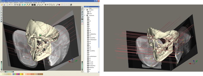
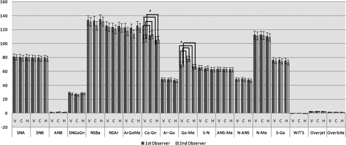
Methods: Pre-treatment cephalometric digital radiographs of 11 patients and their corresponding CBCT images were randomly selected. The digital cephalometric radiographs were traced using Vista Dent OC (GAC International, Inc Bohemia, NY) and by hand. CBCT and Maxilim® (Medicim, Sint-Niklass, Belgium) software were used to generate cephalograms from the CBCT data set that were then linked to the 3D hard-tissue surface representations. In total, 16 cephalometric landmarks were identified and 18 widely used measurements (11 linear and 7 angular) were performed by 2 independent observers. Intraobserver reliability was assessed by calculating intraclass correlation coefficients (ICC), interobserver reliability was assessed with Student t-test and analysis of variance (ANOVA). Mann-Whitney U-tests and Kruskal-Wallis H tests were also used to compare the three methods (P < 0.05).
Results: The results demonstrated no statistically significant difference between interobserver analyses for CBCT-generated cephalograms (P < 0.05), except for Gonion-Menton (Go-Me) and Condylion-Gnathion (Co-Gn). Intraobserver examinations showed low ICCs, which was an indication of poor reproducibility for Go-Me and Sella-Nasion (S-N) in CBCT-generated cephalograms and poor reproducibility for Articulare-Gonion (Ar-Go) in the 2D hand tracing method (P < 0.05). No statistical significance was found for Vista Dent OC measurements (P > 0.05).
Conclusions: Measurements from in vivo CBCT-generated cephalograms from Maxilim® software were found to be similar to conventional images. Thus, owing to higher radiation exposure, CBCT examinations should only be used when the inherent 3D information could improve the outcome of treatment.
Figures
References
-
- Broadbent BH. A new X-ray technique and its application in orthodontics. Angle Orthod 1931;1:45–60
-
- Papadopoulos MA, Jannowitz C, Boettcher P, Henke J, Stolla R, Zeilhofer HF, et al. Three-dimensional fetal cephalometry: an evaluation of the reliability of cephalometric measurements based on three-dimensional CT reconstructions and on dry skulls of sheep fetuses. J Craniomaxillofac Surg 2005;33:229–237 - PubMed
-
- Moreira CR, Sales MA, Lopes PM, Cavalcanti MG. Assessment of linear and angular measurements on three-dimensional cone beam computed tomographic images. Oral Surg Oral Med Oral Pathol Oral Radiol Endod 2009;108:430–436 - PubMed
-
- Chien PC, Parks ET, Eraso F, Hartsfield JK, Roberts WE, Ofner S. Comparison of reliability in anatomical landmark identification using two-dimensional digital cephalometrics and three-dimensional cone beam computed tomography in vivo. Dentomaxillofac Radiol 2009;38:262–273 - PubMed
Publication types
MeSH terms
LinkOut - more resources
Full Text Sources
Medical
Research Materials
