

Case Reports
doi: 10.1259/dmfr/84472023.
Cone beam CT sialography of Stafne bone cavity
Affiliations
- PMID: 22065802
- PMCID: PMC3528154
- DOI: 10.1259/dmfr/84472023
Item in Clipboard
Case Reports
Cone beam CT sialography of Stafne bone cavity
Dentomaxillofac Radiol.
2011 Dec.
Abstract
Stafne bone cavity (SBC) was mostly described as a small oval radiolucency in the posterior mandibular region. To the best of our knowledge, the literature does not contain any report of the use of cone beam CT (CBCT) sialography for the diagnosis of this entity. The aim of this paper is to present a large, irregular and expanded atypical SBC, which made diagnosis difficult. A CBCT sialography was performed to get a definite diagnosis of this rare entity.
Figures
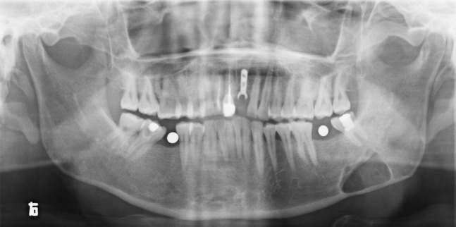
Panoramic radiograph showing well-defined corticated rhomboidal radiolucency in the left mandible. The adjacent third molar was intact and the inferior alveolar canal was overlapped by the lesion
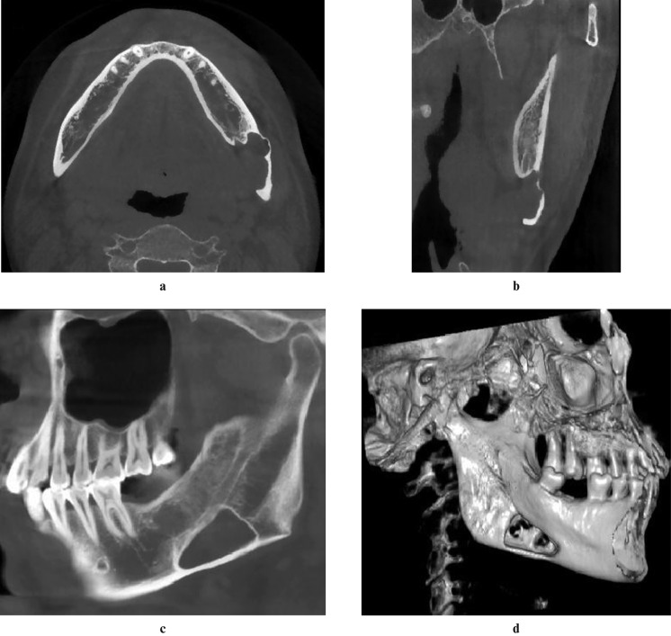
Axial (a), coronal (b) and sagittal (c) sections of cone beam CT (CBCT) showing a large bone cavity with an irregular border. The inner tissue seemed to be continuous with the submandibular gland. The buccal cortex was expanded and perforated in three-dimensional volume rendering processing (d)
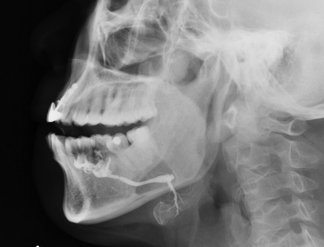
Conventional sialogram showing that some branch ducts arising superiorly from the hilum of the submandibular gland seemed to distribute in the cavity area
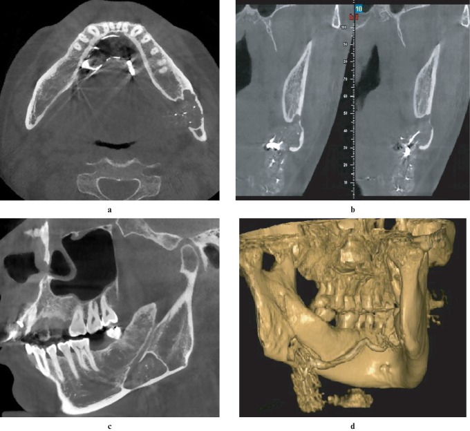
The cavity area was mostly occupied by contrast-filled submandibular gland tissue, and soft tissue of low attenuation was also clearly demonstrated between the cavity wall and gland (a–c). The three-dimensional processing of the cone beam CT (CBCT) sialography shows the spatial relationship of submandibular gland and the bone cavity (d)
References
-
- Stafne E. Bone cavities situated near the angle of the mandible. J Am Dent Assoc 1942;29:1969–1972
-
- Philipsen HP, Takata T, Reichart PA, Sato S, Suei Y. Lingual and buccal mandibular bone depressiones: A review based on 583 cases from a world-wide literature survey, including 69 new cases from Japan. Dentomaxillofac Radiol 2002;31:281–290 - PubMed
-
- Katz J, Chaushu G, Rotstein I. Stafne's bone cavity in the anterior mandible: a possible diagnostic challenge. J Endod 2001;27:304–307 - PubMed
-
- Tsui SH, Chan FF. Lingual mandibular bone defect: case report and review of the literature. Aust Dent J 1994;39:368–371 - PubMed
-
- de Courten A, Kuffer R, Samson J, Lombardi T. Anterior lingual mandibular salivary gland defect (Stafne defect) presenting as a residual cyst. Oral Surg Oral Med Oral Pathol Oral Radiol Endod 2002;94:460–464 - PubMed
