. 2011 Oct 4:5:53.
doi: 10.3389/fnint.2011.00053. eCollection 2011.

Anatomical pathways involved in generating and sensing rhythmic whisker movements

Affiliations

Anatomical pathways involved in generating and sensing rhythmic whisker movements

Laurens W J Bosman et al. Front Integr Neurosci. .

Abstract

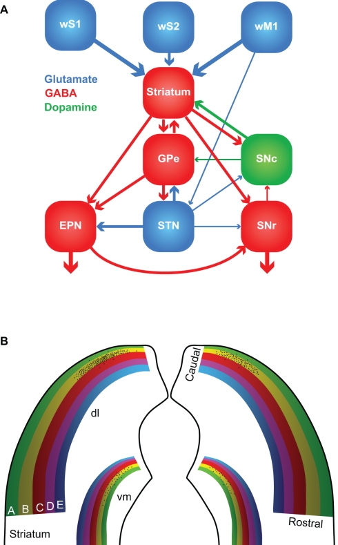
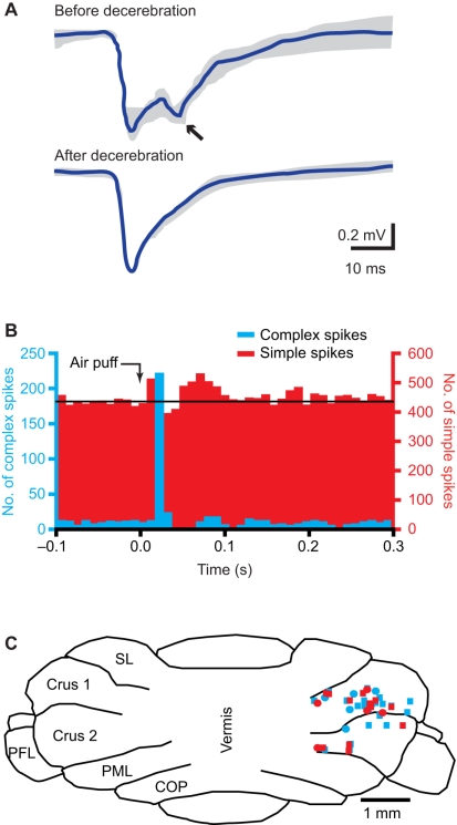
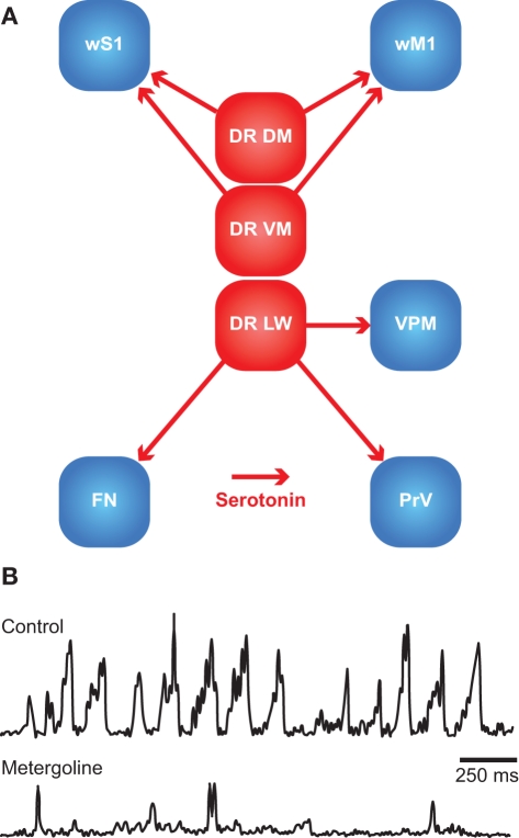
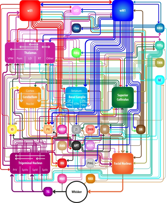
The rodent whisker system is widely used as a model system for investigating sensorimotor integration, neural mechanisms of complex cognitive tasks, neural development, and robotics. The whisker pathways to the barrel cortex have received considerable attention. However, many subcortical structures are paramount to the whisker system. They contribute to important processes, like filtering out salient features, integration with other senses, and adaptation of the whisker system to the general behavioral state of the animal. We present here an overview of the brain regions and their connections involved in the whisker system. We do not only describe the anatomy and functional roles of the cerebral cortex, but also those of subcortical structures like the striatum, superior colliculus, cerebellum, pontomedullary reticular formation, zona incerta, and anterior pretectal nucleus as well as those of level setting systems like the cholinergic, histaminergic, serotonergic, and noradrenergic pathways. We conclude by discussing how these brain regions may affect each other and how they together may control the precise timing of whisker movements and coordinate whisker perception.

Keywords: anatomy; barrel cortex; basal ganglia; cerebellum; follicle–sinus complex; rhythmic movements; sensorimotor integration; vibrissa.


Figures

Figure 1
Figure 1
Whisker movements. (A) Whiskers move rhythmically back-and-forth during exploratory whisking in the rat. The deflection along the rostro-caudal axis is much larger than that on the dorso-ventral axis. The left panel is a schematic drawing of the space in which a whisker can be moved, based on Bermejo et al. (2002). The right panels are reproduced with permission from Hill et al. (2008). (B) Position, velocity, and acceleration of a rat D3 whisker during one whisking cycle on P150 sandpaper. Irregularities in the sandpaper surface cause “slip-stick” movements. Reproduced with permission from Wolfe et al. (2008). (C) Slips can trigger neuronal responses in rat wS1, as shown by a peri-stimulus time histogram of the spike times of a single neuron aligned on the first slips of whisker movements. Reproduced with permission from Jadhav et al. (2009).
Figure 2
Figure 2
Location and structure of the whiskers. (A) The whiskers are organized in rows on the mystacial pad. Mice and rats have five rows of whiskers, as well as four “straddlers” caudal to these rows. Each whisker is associated with an intrinsic capsular muscle [see also (B)]. Extrinsic muscles connect to multiple whiskers. The m. nasolabialis profundis (MNP) consists of two parts, the mediosuperior (PMS) and the medioinferior (PMI) parts, both of which are involved in whisker protraction. The m. nasolabialis and m. maxillolabialis are involved in whisker retraction. The other extrinsic whiskers, including the m. nasolabialis superficialis and the m. buccinatorius, are involved in resizing the entire mystacial pad. The mystacial muscles are almost exclusively innervated by the facial nerve, which leaves the skull via the stylomastoid foramen (SMF). After leaving the SMF, the facial nerve splits up in two streams. The lower stream consists of the rami buccolabialis superior (RBS) and inferior (RBI), which anastomose in the buccal plexus (BP). From the BP all extrinsic and intrinsic whisker muscles are innervated, with the exception of m. nasolabialis, which is innervated by the upper stream, which includes the ramus zygomatico-orbitalis (RZO). (B) Schematic drawing of the follicle–sinus complex (FSC) of the rat. The vibrissa (V) lies within a follicle that is derived from the epidermis and that is surrounded by the glassy membrane (GM). Around the follicle is a blood sinus derived from the dermis, and which is composed of two sinuses: the cavernous sinus (CS), which has numerous collagenous trabeculae, and the ring sinus (RS), which is an open structure. At the bottom of the ring sinus, there is an asymmetric structure of connective tissue: the ringwulst (RW). At the distal end of the ring sinus, the inner conical body (ICB) links the follicle strongly to the capsule (C). Distal to the ICB is the outer conical body (OCB) that contains the sebaceous gland (SG). Intrinsic capsular muscles connect pairs of FSCs. Extrinsic muscles are located just below the skin (m. nasolabialis, MNL and m. maxillolabialis, MML), or at the lower end of the FSC (m. nasolabialis profundus, MNP). The arrows indicate whether contraction of the muscle causes pro- or retraction of the vibrissae. The vibrissae are surrounded by three different types of mechanoreceptors: Merkel cells (MC), lanceolate endings (LE), and free nerve endings (FNE). Mechanoreceptors in the upper part of the FSC are innervated by superficial vibrissal nerves (SVN) and those in the lower part by the deep vibrissal nerve (DVN). In addition, there are some small-caliber fibers at the bottom. The sensory fibers come together with fibers from other parts of the face to form the infraorbital branch of the trigeminal nerve (TN). Blood supply to the FSCs is organized via row arteries (RA) located between the whisker rows, with superficial vibrissal arteries (SVA) supplying the upper parts and deep vibrissal arteries (DVA) the lower parts of the FSCs. The DVA does not directly branch from a RA, but from the anastomozing intervibrissal trunks (IVT). In between the FSCs are intervibrissal arteries (IVA) that supply the skin and hair follicles. The capsular muscles receive their blood from arterioles (PMA) branching from the IVA and directly from the IVT. Venal drainage is organized by intervibrissal veins (IVV) that empty in row veins (RV). (C) Schematic drawings of the follicle of a typical mammalian body hair (left) and of the structure of the blood sinuses of FSCs in different species. A hair follicle lacks a blood sinus and can be moved by contraction of the m. arrector pili. In marsupials and primates, the blood sinus is composed of a single compartment (the cavernous sinus), as illustrated for the tammar wallaby (Macropus eugenii; Marotte et al., 1992) and the rhesus monkey (Van Horn, 1970). Most species, however, have two sinuses: the ring sinus and the cavernous sinus, as illustrated for the rat (Rattus sp.; Ebara et al., 2002) and the Australian water rat (Hydromys chrysogaster; Dehnhardt et al., 1999). Pinnipeds have tricompartite blood sinuses, including an outer cavernous sinus, as illustrated for a sea cow, the Florida manatee (Trichechus manatus latirostris; Reep et al., 2001), and the ringed seal (Phoca hispida; Hyvärinen et al., 2009). Non-whisking species can generally move their vibrissae using m. arrector pili muscles, as indicated for the FSC of the sea cow.
Figure 3
Figure 3
The trigeminal nuclei. (A) The sensory trigeminal nuclei consist of two nuclei, oriented along the antero-posterior axis. The principal nucleus (PrV) is located at the anterior end and the spinal nucleus (SpV) at the posterior site. The SpV can be subdivided into an oral (SpVo), interpolar (SpVi), and caudal part (SpVc). The facial vibrissae project to the ventral part of the trigeminal nuclei. In PrV, SpVc, and the caudal part of SpVi, each vibrissa has its own projection field: a barrelette. The orientation of the barrelettes of the facial macro-vibrissae is indicated schematically. (B) Coronal section of a neonatal mouse brain, showing the location of the barrelettes of the facial macro-vibrissae in the ventral part of SpVi. Following cytochrome oxidase staining, barrelettes appear as dark patches. Note the inverted somatotopy: dorsal vibrissae project to ventral barrelettes. The smaller patches dorsal to the barrelettes of the E-row are the receptive fields of the facial micro-vibrissae. The photomicrograph was kindly provided by Dr. R. S. Erzurumlu.
Figure 4
Figure 4
The trigemino-thalamo-cortical pathways. Schematic drawing of the organization of the barrels in layer 4 of a tangential slice of an adult rat (A), mouse (B), and rabbit (C). Note that the septa are prominent in rats but very small in mice. In adult rabbits, barrels are absent. Instead, the somatotopic representation of the vibrissae is more gradual. (D1) Schematic drawing of a rat brain. The dotted line indicates the recording area for the panels (D2,D3). The red dots indicate the representations of the C2 whisker, and the blue dots those of the E2 whisker in wS1 and wM1. wS2 is partially visible on the extreme left of the recording area. (D2) Voltage-sensitive dye images in urethane-anesthetized mice showing that stimulation of the contralateral C2 whisker initially evokes a very local signal in the C2 barrel of wS1. Consecutively, the signal spreads over the rest of wS1, and also to wM1, and to a lesser extent also to wS2. The time points indicate the time since the onset of whisker deflection, the scale bar the fluorescent signal (blue = weak, white = high). (D3) Idem, but for the E2 whisker. Note that the early responses to the C2 and E2 whiskers are at different locations, but this difference is less obvious during later phases of the response. Panel D is reproduced with permission from Aronoff et al. (2010). (E) Schematic representation of the trigemino-thalamo-cortical pathways discussed in the main text. The arrowheads indicate the termination areas of the axons. Note that (in the cerebral cortex) the postsynaptic cells may have their somata in other layers. The line thickness indicates the relative importance of the pathways. The barreloids in VPM are indicated in an oblique coronal slice, the barrelettes of the trigeminal nuclei in coronal slices. D = dorsal; L, lateral; LD, laterodorsal nucleus of the thalamus; Pom, medial posterior nucleus of the thalamus; PrV, primary trigeminal nucleus; R, rostral; SpVic, caudal part of spinal trigeminal nucleus pars interpolaris; SpVio, oral part of SpVi; SpVo, spinal trigeminal nucleus pars oralis; VPMdm, dorsomedial part of the ventroposterior medial nucleus of the thalamus; VPMh, “head” area of VPM; VPMvl, ventrolateral part of VPM; wM1, whisker motor cortex; wS1, whisker part of primary sensorimotor cortex; wS2, whisker part of secondary sensorimotor cortex.
Figure 5
Figure 5
The basal ganglia. (A) The major connections between the components of the basal ganglia. Most of the input comes from the cerebral cortex (in this case: wS1, wS2, and wM1), and is directed to the striatum. GABAergic medium-spiny neurons of the striatum project to the external part of the globus pallidus (GPe), the entopeduncular nucleus (EPN), and the reticular (SNr) and compact (SNc) parts of the substantia nigra. SNc provides dopaminergic input to the striatum, and the subthalamic nucleus (STN) glutamatergic input to GPe, EPN, and SNr. The main output of the basal ganglia is directed to the thalamus, via GPe and SNr, and the superior colliculus, via SNr. The line thickness indicates the relative importance for the whisker system. (B) Whisker responses in the dorsolateral (dl) striatum follow a loose somatotopy, which is mainly organized according to whisker rows. The black dots indicate schematically the projections of the layer 5 pyramidal cells in the B2 barrel of left wS1. The projection is mainly, but not exclusively, ipsilateral, and largely within the “B row” area in the striatum. There is considerable overlap, however, with the projection areas of other B row whiskers. “Rostral” and “caudal” refer to the positions of the whiskers on the mystacial pad. A smaller and less characterized projection area is also present in the ventromedial (vm) striatum.
Figure 6
Figure 6
The cerebellum. (A) Tactile stimulation of the upper lip evokes a bi-phasic response in the cerebellar cortex, as measured with field potential recordings in the granule cell layer in crus 2 of adult rats. Complete midcollicular decerebration abolished the late phase response, indicating that the late phase response (arrow) is induced by the cerebral cortex, while the early phase is not. Schematic drawing based on Morisette and Bower (1996). (B) Peri-stimulus time histograms of complex spike (blue) and simple spike (red) responses to ipsilateral air puff stimulation of the whiskers in a Purkinje cell in crus 1 of an awake mouse. The complex spike response is uni-phasic, while clear early and late phase simple spike responses can be observed. Reproduced with permission from Bosman et al. (2010). (C) Cross section of the cerebellar cortex, showing the locations where Purkinje cell responses to ipsilateral stimulation of whisker from the C row were observed in crus 1 and crus 2. COP, copula pyramidis; PFL, paraflocculus; PML, paramedian lobule; SL, simple lobule. Modified with permission from Bosman et al. (2010).
Figure 7
Figure 7
The dorsal raphe nuclei as central pattern generator. (A) Schematic overview of the main serotonergic connections from the dorsal raphe nuclei to the brain regions of the whisker system. DR DM, dorsomedial dorsal raphe nucleus; DR LW, lateral wing division of the dorsal raphe nucleus; DR VM, ventromedial dorsal raphe nucleus; FN, facial nucleus; PrV, principal trigeminal nucleus; VPM, medial ventroposterior nucleus; wM1, whisker motor cortex; wS1, barrel cortex. (B) During exploratory whisking, rhythmic whisker movements occur, as shown here by EMG recordings of rat whisker muscles. Application of metergoline, an antagonist for the serotonin receptors 5-HT1 and 5-HT2, in the facial nucleus abolishes the rhythmicity of whisker movements unilaterally at the side of injection. Most likely, the dorsal raphe nuclei are the source of serotonin. This indicates that the dorsal raphe nuclei may act as central pattern generator for whisker movements. Reproduced with permission from Hattox et al. (2003).
Figure 8
Figure 8
Neuronal connections in the whisker system. Many brain regions are involved in controlling the whiskers. Schematic representation of the connections discussed in the main text. Thickness of the arrows corresponds to the robustness of the connection involved (divided among three different levels). Some local connections are indicated, but for the connections between the nuclei of the basal ganglia, see Figure 5A. Amb, ambiguus nucleus; Amg, amygdala; APT, anterior pretectal nucleus; Clau, claustrum; DMN, deep mesencephalic nucleus; DR, dorsal raphe nucleus; EPN, entopeduncular nucleus; GP, globus pallidus; IO, inferior olive; KF–PC, Kölliker-Fuse nucleus and parabrachial complex; LC, locus coeruleus; LD, laterodorsal nucleus; MeV, mesencephalic trigeminal nucleus; NBM, nucleus basalis magnocellularis; NRTP, nucleus reticularis tegmenti pontis; NXII, hypoglossal nucleus; PAG, periaqueductal gray; PN, pontine nucleus; Pom, medial posterior nucleus; PPTg, pedunculopontine tegmental nucleus and the laterodorsal tegmental nucleus; PrV, principal trigeminal nucleus; RF, pontomedullar reticular formation; RN, red nucleus; RT, reticular nucleus; SNc, substantia nigra pars compacta; SNr, substantia nigra pars reticulata; SpVc, spinal trigeminal nucleus pars caudalis; SpVi, spinal trigeminal nucleus pars interpolaris; SpVo, spinal trigeminal nucleus pars oralis; STN, subthalamic nucleus; TG, trigeminal ganglion; TMN, tuberomammillary nucleus; VPM, medial venteroposterior nucleus; wM1, whisker motor cortex; wS1, barrel cortex; wS2, whisker part of the secondary somatosensory cortex; ZI, zona incerta.

References

    1. Ahissar E., Sosnik R., Haidarliu S. (2000). Transformation from temporal to rate coding in a somatosensory thalamocortical pathway. Nature 406, 302–30610.1038/35018568 - DOI - PubMed
    1. Ahl A. S. (1982). Evidence of use of vibrissae in swimming in Sigmodon fulviventer. Anim. Behav. 30, 1203–120610.1016/S0003-3472(82)80211-X - DOI
    1. Ahl A. S. (1986). The role of vibrissae in behavior: a status review. Vet. Res. Commun. 10, 245–26810.1007/BF02213989 - DOI - PubMed
    1. Akaike T. (1992). The tectorecipient zone in the inferior olivary nucleus in the rat. J. Comp. Neurol. 320, 398–41410.1002/cne.903200311 - DOI - PubMed
    1. Alloway K. D. (2008). Information processing streams in rodent barrel cortex: the differential functions of barrel and septal circuits. Cereb. Cortex 18, 979–98910.1093/cercor/bhm138 - DOI - PubMed

LinkOut - more resources