

Acute spontaneous spinal subdural haematoma presenting as paraplegia and complete recovery with non-operative treatment
- PMID: 22065983
- PMCID: PMC3029557
- DOI: 10.1136/bcr.02.2009.1599
Acute spontaneous spinal subdural haematoma presenting as paraplegia and complete recovery with non-operative treatment
Abstract
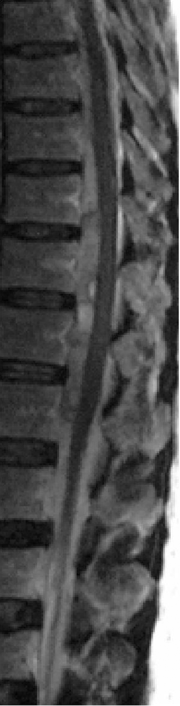
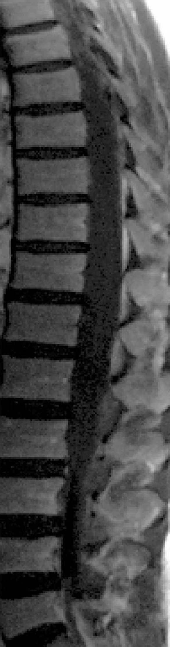
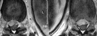
Spontaneous spinal subdural haematoma (SSDH) with no underlying pathology is a very rare condition. Only 20 cases have been previously reported. It can be caused by abnormalities of coagulation, blood dyscrasia, or trauma, underlying neoplasm, and arteriovenous malformation. It occurs most commonly in the thoracic spine and presents with sudden back pain radiating to the arms, legs or trunk, and varying degrees of motor, sensory, and autonomic disturbances. Although the main approach to management is surgical decompression, conservative management is used as well. We report the case of a 57-year-old man who presented with sudden severe low back pain followed by rapid onset of complete paraplegia. Magnetic resonance imaging (MRI) revealed an anterior subdural haematoma from T9 to L1 with cord compression. Corticosteroid treatment was administered. The patient showed substantial clinical improvement after 7 days of bed rest and an intense rehabilitation programme. An MRI scan and a computed tomography angiogram did not reveal any underlying pathology to account for the subdural haematoma.
Figures

Similar articles
-
Acute spinal subdural hematoma: A case report of spontaneous recovery from paraplegia.Medicine (Baltimore). 2020 May;99(19):e20032. doi: 10.1097/MD.0000000000020032. Medicine (Baltimore). 2020. PMID: 32384463 Free PMC article.
-
Acute spontaneous spinal subdural hematoma presenting as paraplegia: a rare case.Spine (Phila Pa 1976). 2007 Oct 1;32(21):E619-22. doi: 10.1097/BRS.0b013e318154c618. Spine (Phila Pa 1976). 2007. PMID: 17906565 Review.
-
Spinal subdural haematoma concurrent with cranial subdural haematoma: Report of two cases and review of literature.Br J Neurosurg. 2010 Oct;24(5):537-41. doi: 10.3109/02688691003656119. Br J Neurosurg. 2010. PMID: 20828301 Review.
-
Spontaneous spinal subdural hematoma: Case study.Am J Crit Care. 2010 Mar;19(2):191-3. doi: 10.4037/ajcc2009982. Epub 2009 Sep 21. Am J Crit Care. 2010. PMID: 19770415
-
[Spontaneous spinal epidural haematoma causing rapid flaccid paraplegia in a healthy 25-year-old patient].Ann Fr Anesth Reanim. 2007 Jun;26(6):608-11. doi: 10.1016/j.annfar.2007.03.010. Epub 2007 Apr 25. Ann Fr Anesth Reanim. 2007. PMID: 17462853 French.
Cited by
-
Spontaneous thoracic ventral spinal subdural hematoma mimicking a tumoral lesion: a case report.J Med Case Rep. 2015 Jun 6;9:132. doi: 10.1186/s13256-015-0562-3. J Med Case Rep. 2015. PMID: 26048171 Free PMC article.
-
Acute, Nontraumatic Spontaneous Spinal Subdural Hematoma: A Case Report and Systematic Review of the Literature.Case Rep Neurol Med. 2017;2017:2431041. doi: 10.1155/2017/2431041. Epub 2017 Dec 26. Case Rep Neurol Med. 2017. PMID: 29441210 Free PMC article.
-
Acute spinal subdural hematoma: A case report of spontaneous recovery from paraplegia.Medicine (Baltimore). 2020 May;99(19):e20032. doi: 10.1097/MD.0000000000020032. Medicine (Baltimore). 2020. PMID: 32384463 Free PMC article.
-
Subarachnoid Hemorrhage and Spinal Subdural Hematoma Due to Acute CSF Hypotension.Neurocrit Care. 2017 Feb;26(1):109-114. doi: 10.1007/s12028-016-0327-x. Neurocrit Care. 2017. PMID: 27660177
References
-
- Russel NA, Benoit BG. Spinal subdural hematoma: a review. Surg Neurol 1983; 20: 133–7 - PubMed
-
- Kyriakides AE, Lalam RK, El Masry WS. Acute spontaneous spinal subdural hematoma presenting as paraplegia. Spine 2007; 32: 619–22 - PubMed
-
- Domenicucci M, Ramieri A, Ciappetta P, et al. Nontraumatic acute spinal subdural haematoma: report of five cases and review of the literature. J Neurosurg 1999; 91: 65–73 - PubMed
-
- Boukobza M, Haddar D, Boissonet M, et al. Spinal subdural haematoma: a study of three cases. Clin Radiol 2001; 56: 475–80 - PubMed
LinkOut - more resources
Full Text Sources