. 2011;6(10):e25660.
doi: 10.1371/journal.pone.0025660. Epub 2011 Oct 17.

Batf3-dependent CD11b(low/-) peripheral dendritic cells are GM-CSF-independent and are not required for Th cell priming after subcutaneous immunization

Affiliations

Batf3-dependent CD11b(low/-) peripheral dendritic cells are GM-CSF-independent and are not required for Th cell priming after subcutaneous immunization

Brian T Edelson et al. PLoS One. 2011.

Abstract

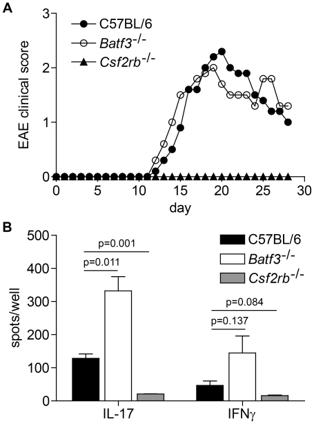
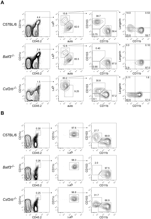
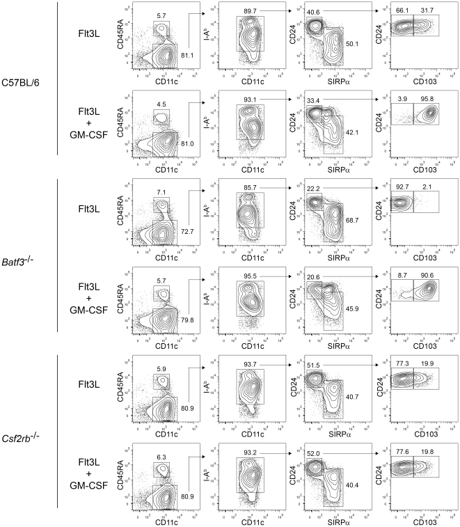
Dendritic cells (DCs) subsets differ in precursor cell of origin, functional properties, requirements for growth factors, and dependence on transcription factors. Lymphoid-tissue resident CD8α(+) conventional DCs (cDCs) and CD11b(low/-)CD103(+) non-lymphoid DCs are developmentally related, each being dependent on FMS-like tyrosine kinase 3 ligand (Flt3L), and requiring the transcription factors Batf3, Irf8, and Id2 for development. It was recently suggested that granulocyte/macrophage colony stimulating factor (GM-CSF) was required for the development of dermal CD11b(low/-)Langerin(+)CD103(+) DCs, and that this dermal DC subset was required for priming autoreactive T cells in experimental autoimmune encephalitis (EAE). Here, we compared development of peripheral tissue DCs and susceptibility to EAE in GM-CSF receptor deficient (Csf2rb(-/-)) and Batf3(-/-) mice. We find that Batf3-dependent dermal CD11b(low/-)Langerin(+) DCs do develop in Csf2rb(-/-) mice, but that they express reduced, but not absent, levels of CD103. Further, Batf3(-/-) mice lacking all peripheral CD11b(low/-) DCs show robust Th cell priming after subcutaneous immunization and are susceptible to EAE. Our results suggest that defective T effector priming and resistance to EAE exhibited by Csf2rb(-/-) mice does not result from the absence of dermal CD11b(low/-)Langerin(+)CD103(+) DCs.


Conflict of interest statement

Competing Interests: The authors have declared that no competing interests exist.

Figures

Figure 1
Figure 1. Batf3 −/− mice are susceptible to EAE.
(A) Clinical course of EAE in C57BL/6 mice, Batf3 −/− mice (C57BL/6 background), and Csf2rb −/− mice. Data are combined from two experiments (n = 10 mice per group). Points represent the mean clinical score. For clarity, error bars are not displayed. (B) ELISPOT assays on popliteal lymph node cells at day 7 following footpad immunization (MOG35–55 peptide in CFA) and restimulation with MOG35–55 peptide. Data are from one of two similar experiments (n = 3 per group). Mean ± SEM, p values by unpaired student's t test.
Figure 2
Figure 2. Batf3-dependent dermal CD11blow/−Langerin+ DCs develop in Csf2rb −/− mice, but lack expression of CD103.
(A) FACS analysis of SDLN (inguinal) DCs from C57BL/6, Batf3 −/− (C57BL/6 background), and Csf2rb −/− mice. Left plots are gated on CD11chigh cells. The second column is gated on migratory (DEC205+CD8α) DCs. The third, fourth, and fifth columns are gated on Langerin+ migratory DCs. Numbers represent the percentage of cells within the indicated gates. MFI are shown for the indicated gates. Data are representative of eight to ten individual inguinal lymph nodes from four to five mice per genotype over two experiments. (B) FACS analysis of SDLN (inguinal) DCs from C57BL/6, Batf3 −/−, and Csf2rb −/− mice. Left plots are gated on CD11chigh cells. Middle plots are gated on migratory (DEC205+CD8α) DCs. Right plots are gated on all Langerin+ migratory DCs, independent of expression of CD11b. Numbers represent the percentage of cells within the indicated gates. Data are representative of inguinal lymph nodes from at least five mice per genotype for all stains except CD24, which has been performed on one mouse per genotype. (C) Absolute cell numbers per individual inguinal lymph nodes calculated from the FACS analysis in (A). Langerhans cells were gated as Langerin+CD11b+EpCAMhigh DCs, and dermal Langerin+ DCs were gated as Langerin+CD11blow/−EpCAMmid DCs. Horizontal bars represent the geometric mean; p values by unpaired student's t test. (D) Clinical course of EAE in 129S6/SvEv mice and Batf3 −/− mice (129S6/SvEv background). Data are from one of three similar experiments (n = 5 mice per group). Points represent the mean clinical score. For clarity, error bars are not displayed.
Figure 3
Figure 3. GM-CSF controls the level of CD103 expression on Batf3-dependent CD11blow/− peripheral DCs.
(A) FACS analysis of lung DCs from C57BL/6, Batf3 −/− (C57BL/6 background), and Csf2rb −/− mice. Left plots are gated on live, single cells. The second column of plots is gated on CD11c+CD45.2+ cells, consisting of a mixture of lung macrophages and DCs. Note that in Csf2rb −/− mice, lung macrophages lack CD11c expression , and therefore this gate includes only DCs. The third and fourth columns of plots are gated on I-Abhigh autofluorescencelow DCs. Numbers represent the percentage of cells within the indicated gates. Data are representative of six individual lungs from three mice per genotype over two experiments. (B) FACS analysis of kidney DCs from C57BL/6, Batf3 −/− (C57BL/6 background), and Csf2rb −/− mice. Left plots are gated on live, single cells. The second column of plots is gated on CD11chighCD45.2+ cells, consisting of DCs. The third column of plots is gated on CD11chighI-Ab+ DCs. Numbers represent the percentage of cells within the indicated gates. Data are representative of six individual kidneys from three mice per genotype over two experiments.
Figure 4
Figure 4. GM-CSF induces CD103 expression on Flt3L-derived CD8α-equivalent DCs.
FACS analysis of developing DCs in bone marrow cells from C57BL/6, Batf3 −/− (C57BL/6 background), and Csf2rb −/− mice cultured with Flt3L for 9 days, with or without the addition of GM-CSF to the culture on day 7. The left column is gated on live single cells, the second on CD11c+CD45RA cDCs, the third on I-Ab+ cDCs, and the fourth on CD24highSirpαlow cells. Numbers represent the percentage of cells within the indicated gates. Data are representative of bone marrow cells from two mice per genotype over two experiments.

References

    1. Hamilton JA. Colony-stimulating factors in inflammation and autoimmunity. Nat Rev Immunol. 2008;8:533–544. - PubMed
    1. McQualter JL, Darwiche R, Ewing C, Onuki M, Kay TW, et al. Granulocyte macrophage colony-stimulating factor: a new putative therapeutic target in multiple sclerosis. J Exp Med. 2001;194:873–882. - PMC - PubMed
    1. Cook AD, Braine EL, Campbell IK, Rich MJ, Hamilton JA. Blockade of collagen-induced arthritis post-onset by antibody to granulocyte-macrophage colony-stimulating factor (GM-CSF): requirement for GM-CSF in the effector phase of disease. Arthritis Res. 2001;3:293–298. - PMC - PubMed
    1. Sonderegger I, Iezzi G, Maier R, Schmitz N, Kurrer M, et al. GM-CSF mediates autoimmunity by enhancing IL-6-dependent Th17 cell development and survival. J Exp Med. 2008;205:2281–2294. - PMC - PubMed
    1. Ponomarev ED, Shriver LP, Maresz K, Pedras-Vasconcelos J, Verthelyi D, et al. GM-CSF production by autoreactive T cells is required for the activation of microglial cells and the onset of experimental autoimmune encephalomyelitis. J Immunol. 2007;178:39–48. - PubMed

Publication types

MeSH terms