. 2011;6(10):e26839.
doi: 10.1371/journal.pone.0026839. Epub 2011 Oct 31.

Pathobiological implications of MUC16 expression in pancreatic cancer

Affiliations

Pathobiological implications of MUC16 expression in pancreatic cancer

Dhanya Haridas et al. PLoS One. 2011.

Abstract

MUC16 (CA125) belongs to a family of high-molecular weight O-glycosylated proteins known as mucins. While MUC16 is well known as a biomarker in ovarian cancer, its expression pattern in pancreatic cancer (PC), the fourth leading cause of cancer related deaths in the United States, remains unknown. The aim of our study was to analyze the expression of MUC16 during the initiation, progression and metastasis of PC for possible implication in PC diagnosis, prognosis and therapy. In this study, a microarray containing tissues from healthy and PC patients was used to investigate the differential protein expression of MUC16 in PC. MUC16 mRNA levels were also measured by RT-PCR in the normal human pancreatic, pancreatitis, and PC tissues. To investigate its expression pattern during PC metastasis, tissue samples from the primary pancreatic tumor and metastases (from the same patient) in the lymph nodes, liver, lung and omentum from Stage IV PC patients were analyzed. To determine its association in the initiation of PC, tissues from PC patients containing pre-neoplastic lesions of varying grades were stained for MUC16. Finally, MUC16 expression was analyzed in 18 human PC cell lines. MUC16 is not expressed in the normal pancreatic ducts and is strongly upregulated in PC and detected in pancreatitis tissue. It is first detected in the high-grade pre-neoplastic lesions preceding invasive adenocarcinoma, suggesting that its upregulation is a late event during the initiation of this disease. MUC16 expression appears to be stronger in metastatic lesions when compared to the primary tumor, suggesting a role in PC metastasis. We have also identified PC cell lines that express MUC16, which can be used in future studies to elucidate its functional role in PC. Altogether, our results reveal that MUC16 expression is significantly increased in PC and could play a potential role in the progression of this disease.


Conflict of interest statement

Competing Interests: The authors have declared that no competing interests exist.

Figures

Figure 1
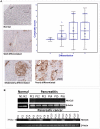
Figure 1. The expression of MUC16 in normal (N) and pancreatic cancer (PC) tissues by immunohistochemistry.
The tissue sections were obtained from ACCUMAX in the form of an array and were stained with anti-MUC16 monoclonal antibody. The stained sections were observed under the microscope and the immunoreactivity was judged by the intensity and spread of the dark stain. Anti-MUC16 antibody showed no staining in the normal pancreas tissue (both ducts and acini) while a strong staining was observed in the cancerous tissues. (A) Box plot representing the score of MUC16 across the various grades of PC. From the box plot we observed the immunoreactivity to be higher in the poorly (D) and moderately differentiated (C) tissues in comparison to well differentiated (B) tissues. The normal pancreas (A) tissue is also negative. Representative sections demonstrating MUC16 expression in normal pancreatic ducts and various grades of invasive adenocarcinoma are shown. Note the predominant membrane staining of MUC16 in PC. (B) Expression studies of MUC16 in normal, pancreatitis and pancreatic cancer tissues by RT-PCR. RT-PCR was performed on mRNA isolated from normal human pancreatic tissue (N1, N2), human pancreatitis tissue (Pt1–Pt6) and human pancreatic cancer tissue (PC1–PC17). No amplification was observed in the normal tissues but amplification was observed in the pancreatic cancer and pancreatitis tissues. Actin was used as an internal control.
Figure 2
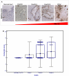
Figure 2. Expression of MUC16 in PanIN lesions and normal ducts.
(A) MUC16 expression was evaluated in tissues containing both the normal pancreas, and adjacent dysplastic lesions. While MUC16 expression was weak in the low-grade, early stage PanIN lesions (PanIN I), it progressively increases with increasing dysplasia with the highest expression observed in high-grade dysplasia (PanIN III) and PC. Note the predominant membrane staining of MUC16 in all grades of PanINs (Original magnification ×200). (B) Box plot representing the composite score of MUC16 across the different grades of PanIN lesions and normal ducts. From the box plot we observe that MUC16 is strongly expressed in PanIN II and III when compared to PanIN I.
Figure 3
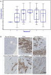
Figure 3. Expression of MUC16 in matched primary and metastatic pancreatic cancer tissues.
To investigate the alteration in MUC16 expression with progression, we investigated the expression of MUC16 in matched primary pancreatic cancer and metastasis to either the lung, lymph node, liver or the omentum/diaphragm. In (A) while the expression of MUC16 was higher in metastatic PC than in the primary tumor, this was not significant. A- Normal pancreas; B- pancreatic cancer; C- Liver metastasis; D- Lung metastasis; E- Lymph Node metastasis; F- Omentum/Diaphragm metastasis. Further, (B) shows that in the same patient, the non-neoplastic ducts were negative, while there was a strong expression of MUC16 in the primary pancreatic tumor and this was maintained even in the metastasis.
Figure 4
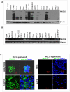
Figure 4. Expression of MUC16 in a panel of PC cell lines using western blot, RT-PCR and confocal studies.
(A) Western blot analysis of MUC16 expression in PC cell lines. Protein lysates from eighteen PC cell lines were resolved on a 2% SDS-agarose gel. MUC16 expression was observed in DanG, HPAC, SU86.86, Colo357, CD18/HPAF, Capan1 and T3M4 cell lines. β-actin was used as an internal control (B) RT-PCR analysis of MUC16 expression in various PC cell lines. Actin was used as an internal control. (C) Immunofluorescence studies of two cell lines (CD18 and Capan1) that expresses MUC16 and two cell lines (CD11 and SUIT2) that do not express MUC16.

References

    1. Jemal A, Siegel R, Xu J, Ward E. Cancer statistics, 2010. CA Cancer J Clin. 2010;60:277–300. - PubMed
    1. Kloppel G, Lingenthal G, von BM, Kern HF. Histological and fine structural features of pancreatic ductal adenocarcinomas in relation to growth and prognosis: studies in xenografted tumours and clinico-histopathological correlation in a series of 75 cases. Histopathology. 1985;9:841–856. - PubMed
    1. Andrianifahanana M, Moniaux N, Batra SK. Regulation of mucin expression: mechanistic aspects and implications for cancer and inflammatory diseases. Biochim Biophys Acta. 2006;1765:189–222. - PubMed
    1. Bafna S, Kaur S, Batra SK. Membrane-bound mucins: the mechanistic basis for alterations in the growth and survival of cancer cells. Oncogene. 2010;29:2893–2904. - PMC - PubMed
    1. Chaturvedi P, Singh AP, Batra SK. Structure, evolution, and biology of the MUC4 mucin. FASEB J. 2008;22:966–981. - PMC - PubMed

Publication types

MeSH terms