Cystic lymphangioma of the pancreas mimicking pancreatic pseudocyst
- PMID: 22066085
- PMCID: PMC3205361
- DOI: 10.4174/jkss.2011.80.Suppl1.S55
Cystic lymphangioma of the pancreas mimicking pancreatic pseudocyst
Abstract
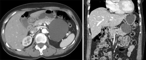
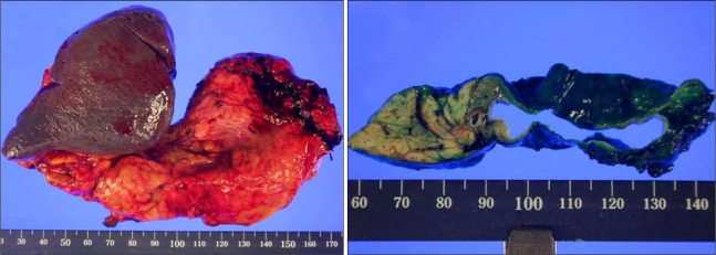
Lymphangiomas are rare congenital benign tumors arising from the lymphatic system, and are mostly encountered in the neck and axillary regions of pediatric patients (95%). Lymphangioma of the pancreas is extremely rare accounting for less than 1% of these tumors. We report here on a case of pancreatic cystic lymphangioma. A 54-year-old woman presented with intermittent postprandial abdominal discomfort and radiating back pain. Abdominal computed tomography scan revealed 8 × 6.5 cm hypodense cystic mass arising from the tail of the pancreas without septa or solid component. The initial impression was a pancreatic pseudocyst. The patient underwent distal pancreatectomy with splenectomy. The histopathologic and immunohistochemical study helped make the diagnosis of a pancreatic cystic lymphangioma. Herein, we report a case of pancreatic cystic lymphangioma mimicking pancreatic pseudocyst and review the relevant medical literature.
Keywords: Cystic lymphangioma; Pancreas; Pseudocyst; Surgical excision.
Conflict of interest statement
No potential conflict of interest relevant to this article was reported.
Figures

References
-
- Koenig TR, Loyer EM, Whitman GJ, Raymond AK, Charnsangavej C. Cystic lymphangioma of the pancreas. AJR Am J Roentgenol. 2001;177:1090. - PubMed
-
- Igarashi A, Maruo Y, Ito T, Ohsawa K, Serizawa A, Yabe M, et al. Huge cystic lymphangioma of the pancreas: report of a case. Surg Today. 2001;31:743–746. - PubMed
-
- Casadei R, Minni F, Selva S, Marrano N, Marrano D. Cystic lymphangioma of the pancreas: anatomoclinical, diagnostic and therapeutic considerations regarding three personal observations and review of the literature. Hepatogastroenterology. 2003;50:1681–1686. - PubMed
-
- Lyngdoh TS, Konsam R, Th B, Marak B. Giant cystic lymphangioma of pancreas. ANZ J Surg. 2008;78:673–674. - PubMed
-
- Schneider G, Seidel R, Altmeyer K, Remberger K, Pistorius G, Kramann B, et al. Lymphangioma of the pancreas and the duodenal wall: MR imaging findings. Eur Radiol. 2001;11:2232–2235. - PubMed
Publication types
LinkOut - more resources
Full Text Sources