Review
. 2012 Mar 15;16(6):543-66.
doi: 10.1089/ars.2011.4119. Epub 2012 Jan 6.

Mechanisms of altered redox regulation in neurodegenerative diseases--focus on S--glutathionylation

Affiliations
Review

Mechanisms of altered redox regulation in neurodegenerative diseases--focus on S--glutathionylation

Elizabeth A Sabens Liedhegner et al. Antioxid Redox Signal. .

Abstract

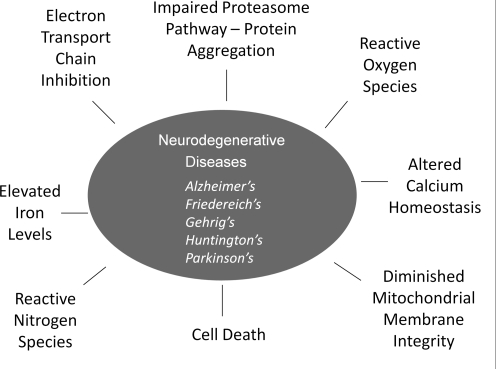
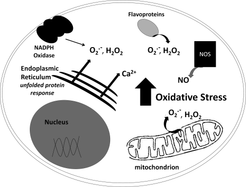
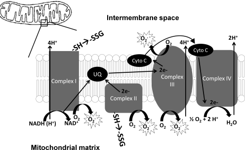
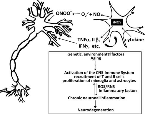
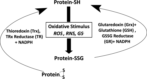
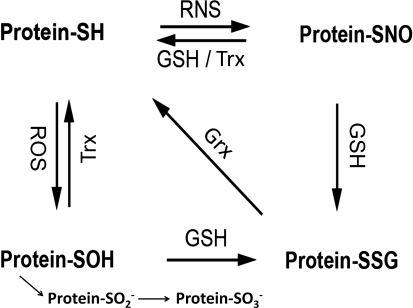
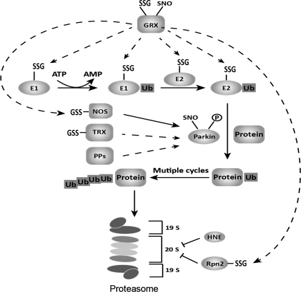
Significance: Neurodegenerative diseases are characterized by progressive loss of neurons. A common feature is oxidative stress, which arises when reactive oxygen species (ROS) and/or reactive nitrogen species (RNS) exceed amounts required for normal redox signaling. An imbalance in ROS/RNS alters functionality of cysteines and perturbs thiol-disulfide homeostasis. Many cysteine modifications may occur, but reversible protein mixed disulfides with glutathione (GSH) likely represents the common steady-state derivative due to cellular abundance of GSH and ready conversion of cysteine-sulfenic acid and S-nitrosocysteine precursors to S-glutathionylcysteine disulfides. Thus, S-glutathionylation acts in redox signal transduction and serves as a protective mechanism against irreversible cysteine oxidation. Reversal of protein-S-glutathionylation is catalyzed specifically by glutaredoxin which thereby plays a critical role in cellular regulation. This review highlights the role of oxidative modification of proteins, notably S-glutathionylation, and alterations in thiol homeostatic enzyme activities in neurodegenerative diseases, providing insights for therapeutic intervention.

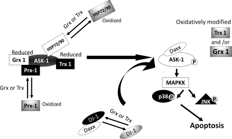
Recent advances: Recent studies show that dysregulation of redox signaling and sulfhydryl homeostasis likely contributes to onset/progression of neurodegeneration. Oxidative stress alters the thiol-disulfide status of key proteins that regulate the balance between cell survival and cell death.

Critical issues: Much of the current information about redox modification of key enzymes and signaling intermediates has been gleaned from studies focused on oxidative stress situations other than the neurodegenerative diseases.

Future directions: The findings in other contexts are expected to apply to understanding neurodegenerative mechanisms. Identification of selectively glutathionylated proteins in a quantitative fashion will provide new insights about neuropathological consequences of this oxidative protein modification.


Figures

FIG. 1.
FIG. 1.
Schematic summary of the neurodegenerative diseases and their common molecular features. The five common neurodegenerative diseases are listed along with their common feature of neuronal cell death that is mediated by oxidative and nitrosative stress conditions and disruption of vital functions, including mitochondrial respiration and proteasomal degradation of altered proteins.
FIG. 2.
FIG. 2.
Schematic representation of cellular sources of reactive species. Shown schematically are the primary sources of reactive oxgen species (ROS) and reactive nitrogen species (RNS), including NADPH oxidase (NOX) at the cellular membrane, oxidases of the endoplasmic reticulum, flavoenzymes (cytosol and ER), nitric oxide synthetase (NOS), and the mitochondria.
FIG. 3.
FIG. 3.
Mitochondrial generation of ROS. A schematic representation of the electron transport chain (ETC) is depicted here. Complex I, Complex II, and Complex III have all been implicated as generators of superoxide, leading to hydrogen peroxide formation.
FIG. 4.
FIG. 4.
Activated microglia promote inflammatory assault on neurons. A schematic representation of an activated microglial cell and a neuron is depicted here. Microglia, the resident macrophages of the central nervous system (CNS) are activated by and produce cytokines and RNS and ROS that mediate external oxidative stress impacting the neuronal cells. Different insults, such as genetic and environmental factors or aging, affecting the CNS can lead to activation of the CNS–immune system, including lymphocyte recruitment, microglia activation, and astrocyte proliferation. These may in turn induce the production of reactive oxygen species (ROS) or reactive nitrogen species (RNS) and drive the expression of inflammatory factors. Those inflammatory mediators can influence the fate of neurons and stimulate the CNS-immune cells to amplify proinflammatory signals and induce neurotoxic effects. Uncontrolled or chronic inflammation can result in loss of neurons and progression of neurodegenerative diseases.
FIG. 5.
FIG. 5.
Glutaredoxin and thioredoxin systems contribute synergistically to sulfhydryl homeostasis. Besides scavenging of reactive species, the other important aspect of thiol homeostasis is repair of sulfhydryl modifications. This function is performed by the thiol-disulfide oxidoreductase (TDOR) enzyme systems. Glutaredoxin (Grx, also known as thioltransferase), coupled to GSH and GSSG reductase, and the thioredoxin (Trx)–thioredoxin reductase system catalyze disulfide reduction and reactivation of oxidatively-modified sulfhydryl proteins. The Trx system favors reduction of intramolecular disulfides via a Trx-(S-S) intramolecular disulfide intermediate, and it is indiscriminate with mixed disulfides substrates. Grx is highly selective for glutathione-containing mixed disulfides (i.e., protein-SSG). Thus, reversible protein–SSG formation by Grx may protect vital proteins from irreversible damage and serve as a regulatory mechanism. As described in the text, many of the proteins characteristically associated with neurodegenerative diseases are subject to oxidative modification and potential regulation via this mechanism.
FIG. 6.
FIG. 6.
Interconversion of forms of modified cysteine residues on proteins. According to the relative reactivity and relative abundance of ROS, RNS, and GSH (GSSG) in cells under conditions of redox signaling or oxidative stress, the expected sequence of events is initial formation of protein-SOH which is readily converted to Protein-SSG; or initial formation of Protein-SNO which is readily converted to Protein-SSG. Thus, Grx plays a key role in determining the steady-state level of Protein-SSG under various conditions (64). Certain proteins that sequester the S-NO or S-OH moieties may be resistant to conversion to Protein-SSG. Also, Trx is reported to catalyze denitrosylation (20). In addition, the S-OH moiety may undergo additional oxidation to sulfinic (-SO2H) and sulfonic (-SO3H) acid forms which are essentially irreversible (see text).
FIG. 7.
FIG. 7.
Schematic representation of potential modes of ASK1 activation under oxidative stress. On the left, ASK1 is represented in its inactive form in the cytosol, bound to some of its multiple negative regulators. Upon oxidative stimulus (e.g., inflammatory mediators, L-DOPA treatment), these negative regulators become oxidized or otherwise modified (e.g., glutathionylation, dopaquinone adduction) and dissociate from ASK1. Concomitantly, ASK 1 is activated wherein autophosphorylated (activated) ASK1 initiates a phosphorylation cascade of downstream mediators, which induces apoptosis (shown at the right). L-DOPA treatment has been shown to cause loss of Grx1 and Trx1 activities (154), which would impede reduction of other oxidized negative regulators of ASK1, resulting in prolonged ASK1 activation. Details of this model, including the potential involvement of Daxx and DJ-1, are presented in the text.
FIG. 8.
FIG. 8.
Regulation of intracellular ionized calcium. This scheme provides a representation of the various calcium channels and their effects on intracellular ionized calcium (increase or decrease) and identifies those which have been reported to be modified by S-glutathionylation. Ca2+ flux is a critical and tightly controlled aspect of normal cell activity. Dysregulation of intracellular ionized calcium concentration occurs with aging and oxidative stress, resulting in decreased cell viability. Several transporters that control intracellular calcium are subject to functional change by oxidation, [e.g., reversible S-glutathionylation (see text)]. Ca2+ flux is regulated by several transporters including two calcium ATPases associated with the smooth endoplasmic reticulum and the plasma membrane (SERCA and PMCA), two receptors located on the SER membrane (RyR, and IP3 receptor), and two transporters located within mitochondria (MPTP and MCT). The two Ca2+ ATPases extrude cytosolic calcium into sequestered compartments: SERCA moves most of the Ca2+ from the cytoplasm into the SER, while the plasma membrane calcium ATPase (PMCA) pumps Ca2+ into the extracellular milieu. RyR and IP3R release Ca2+ from the SER into the cytosol. The mitochondrial calcium transporter (MCT) promotes calcium reuptake, while the mitochondrial permeability pore (MPP) extrudes calcium.
FIG. 9.
FIG. 9.
Post-translational redox modifications in the ubiquitin–proteasome system. Various oxidative modifications, mainly Cys-based (glutathionylation, nitrosylation), have been identified in proteins involved in the ubiquitin proteasome system (UPS). Previous studies have defined a putative role for each modification, for example, S-nitrosylation of Parkin (Parkin-SNO) triggered by generation of NO from nitric oxide synthase (NOS) can cause neuronal cell death through the accumulation of aberrant proteins; S-glutathionylation of ubiquitin ligase E1, 2 and 20S proteasome regulator Rpn2 can downregulate the intracellular proteasome degradation system. Recently, NOS itself was found to undergo glutathionylation, altering its function from NO production to superoxide production (see text). The Cys-based modifications are controlled by the oxidoreductases glutaredoxin (Grx) and thioredoxin (Trx), which mediate deglutathionylation and denitrosylation, respectively. Furthermore, human Grx and Trx have been reported to undergo glutathionylation or nitrosylation themselves, leading to inactivation in a context-dependent manner. It is still unclear whether the –SNO and –SSG modifications work in concert and if the modifications can influence each other. It appears that such modifications at various points in the UPS pathway may determine the accumulation of misfolded or aberrant proteins in neurodegeneration.

References

    1. Adachi T. Pimentel DR. Heibeck T. Hou X. Lee YJ. Jiang B. Ido Y. Cohen RA. S-glutathiolation of Ras mediates redox-sensitive signaling by angiotensin II in vascular smooth muscle cells. J Biol Chem. 2004;279:29857–29862. - PubMed
    1. Adachi T. Weisbrod RM. Pimentel DR. Ying J. Sharov VS. Schoneich C. Cohen RA. S-Glutathiolation by peroxynitrite activates SERCA during arterial relaxation by nitric oxide. Nat Med. 2004;10:1200–1207. - PubMed
    1. Adam-Vizi V. Production of reactive oxygen species in brain mitochondria: Contribution by electron transport chain and non-electron transport chain sources. Antioxid Redox Signal. 2005;7:1140–1149. - PubMed
    1. Aksenov MY. Aksenova MV. Butterfield DA. Geddes JW. Markesbery WR. Protein oxidation in the brain in Alzheimer's disease. Neuroscience. 2001;103:373–383. - PubMed
    1. Akterin S. Cowburn RF. Miranda-Vizuete A. Jimenez A. Bogdanovic N. Winblad B. Cedazo-Minguez A. Involvement of glutaredoxin-1 and thioredoxin-1 in beta-amyloid toxicity and Alzheimer's disease. Cell Death Differ. 2006;13:1454–1465. - PubMed

Publication types