. 2012 Jan;220(1):3-12.
doi: 10.1111/j.1469-7580.2011.01452.x. Epub 2011 Nov 8.

Denervation affects regenerative responses in MRL/MpJ and repair in C57BL/6 ear wounds

Affiliations

Denervation affects regenerative responses in MRL/MpJ and repair in C57BL/6 ear wounds

Gemma Buckley et al. J Anat. 2012 Jan.

Abstract

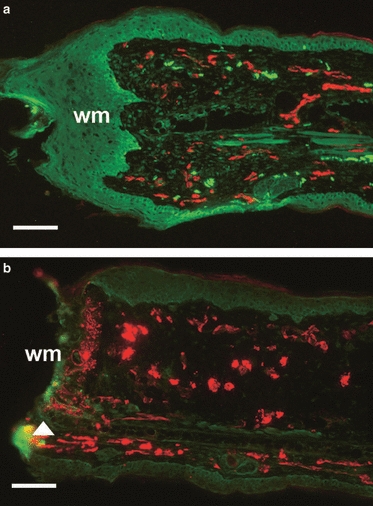
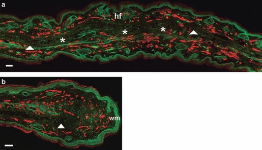
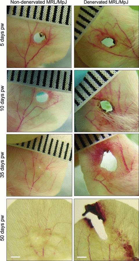
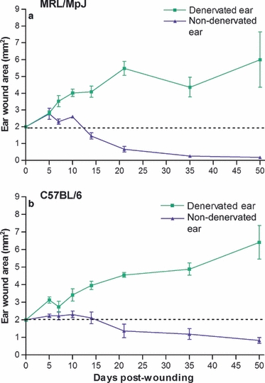
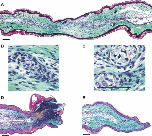
The MRL/MpJ mouse displays the rare ability amongst mammals to heal injured ear tissue without scarring. Numerous studies have shown that the formation of a blastema-like structure leads to subsequent tissue regeneration in this model, indicating many parallels with amphibian limb regeneration and mammalian embryogenesis. We have recently shown that the MRL/MpJ mouse also possesses an enhanced capacity for peripheral nerve regeneration within the ear wound. Indeed, nerves are vital for the initial phase of blastema formation in the amphibian limb. In this study we investigated the capacity for wound regeneration in a denervated ear. The left ears of MRL/MpJ mice and C57BL/6 (a control strain known to have a poorer regenerative capacity) were surgically denervated at the base via an incision and nerve transection, immediately followed by a 2-mm ear punch wound. Immunohistochemical analysis showed a lack of neurofilament expression in the denervated ear wound. Histology revealed that denervation prevented blastema formation and chrondrogenesis, and also severely hindered normal healing, with disrupted re-epithelialisation, increasing wound size and progressive necrosis towards the ear tip. Denervation of the ear obliterated the regenerative capacity of the MRL/MpJ mouse, and also had a severe negative effect on the ear wound repair mechanisms of the C57BL/6 strain. These data suggest that innervation may be important not only for regeneration but also for normal wound repair processes.


Figures

Fig. 1
Fig. 1
Re-innervation and vascularisation of denervated vs. non-denervated MRL/MpJ ear wounds 7 days post-wounding. Immunohistochemistry assessed the presence of pan-neurofilament (FITC) and CD31 (TRITC) in both non-denervated ear wounds (a) and denervated ear wounds (b) in the MRL/MpJ mouse 7 days post-wounding. (a) Section from the centre of the regenerating ear showing the presence of nerves and blood vessels advancing beyond the cut edge of the cartilage (arrow) and a thickened wound epithelium at the wound margin (wm). (b) Section from the denervated ear showing the denervation procedure was successful as no nerves were present and the blood vessels appear dilated. Note the speckled staining of CD31 (arrow), indicating disintegration of vessels at the necrosing wound margin (wm). Scale bars: 100 μm.
Fig. 2
Fig. 2
Re-innervation and vascularisation of denervated vs. innervated non-denervated MRL/MpJ ear wounds 50 days post-wounding. Immunohistochemistry was used to detect the presence of neurofilament (FITC) and CD31 (TRITC) in both non-denervated ear wounds (a) and denervated ear wounds (b) in the MRL/MpJ mouse 50 days post-wounding. (a) Section taken from regenerating ear tissue showing cartilage island formation (*), regenerated hair follicles (hf) beyond the original wound margin (wm; arrows). (b) Section from proximal wound margin of a denervated ear showing re-vascularisation, but only a small influx of regenerating nerves between the cut edge of cartilage (arrow) and the wound margin (wm). Scale bars: 100 μm.
Fig. 3
Fig. 3
Macroscopic photographs of MRL/MpJ ear wound healing in a non-denervated (left) vs. denervated ear (right) 5–50 days post-wounding (pw). MRL/MpJ ear wounds photographed at days 5, 10, 35 and 50 post-wounding (representative examples shown). The non-denervated wound re-epithelialised and regenerated to nearly close the wound hole by day 50. The denervated ear wound became increasingly necrotic, failed to re-epithelialise distally, increased in size and extended out toward the ear tip. Ruler graduations: 1 mm; scale bars: 2 mm.
Fig. 4
Fig. 4
Effect of denervation on wound area. Both denervated and non-denervated ears were wounded with a 2-mm clinical biopsy punch. Denervation of ear wounds caused a significant increase in punch hole area (*P ≤0.05) compared with non-denervated ear wounds from day 7 post-wounding in MRL/MpJ (a) and day 10 in C57BL/6 (b). Ear hole diameter was measured at intervals of 5, 7, 10, 14, 21, 35 and 50 days post-wounding. Image pro plus software was used to calculate wound area. Data shown represent means ± SEM, MRL/MpJ; n = 6, C57BL/6; n = 6 for each time point.
Fig. 5
Fig. 5
Histology of the denervated and non-denervated MRL/MpJ ear wounds 50 days post-wounding. Masson's Trichrome stained tissue sections. Non-denervated ear: (A) section through the centre of the regenerating wound displaying collagen deposition, epithelial fusion of opposing wound margins, and chondrogenesis with the formation of numerous cartilage islands (magnified in B and C). Denervated ear: (D) section through the distal edge of the wound at the necrosing ear tip, displaying severe regression of the wound with necrosis and failure to re-epithelialise; (E) section through the proximal wound edge showing re-epithelialisation, collagen deposition and proliferation at the cartilage stump (arrow). However, no characteristic features of a regenerative blastema-like structure were observed in the denervated MRL/MpJ ear wound. Scale bars: 100 μm.
Fig. 6
Fig. 6
Macroscopic photographs and histology of the denervated and non-denervated C57BL/6 ear wound 50 days post-wounding. (a) Macroscopic photograph of the non-denervated C57BL/6 wound 50 days post-wounding showing limited wound closure (figures and arrows correspond to cross-sections sampled for histology displayed below). Masson's Trichrome stained tissue sections; (a1) through distal wound margin and (a2) through proximal wound margin showing a bulbous aggregate of cartilage. (b) Photograph of the C57BL/6 denervated ear wound 50 days post-wounding showing extension of wound area towards the ear tip (b1) through the distal tip of the ear wound displaying extensive necrosis and failure to re-epithelialise, and (b2) through the proximal ear wound margin showing re-epithelialisation, but no evidence of cartilage formation or dermal extension. Scale bars: 100 μm.

References

    1. Barker AR, Rosson GD, Dellon AL. Wound healing in denervated tissue. Ann Plast Surg. 2006;57:339–342. - PubMed
    1. Beare AH, Metcalfe AD, Ferguson MW. Location of injury influences the mechanisms of both regeneration and repair within the MRL/MpJ mouse. J Anat. 2006;209:547–559. - PMC - PubMed
    1. Bedelbaeva K, Snyder A, Gourevitch D, et al. Lack of p21 expression links cell cycle control and appendage regeneration in mice. PNAS. 2010;30:107. - PMC - PubMed
    1. Brockes JP, Kumar A. Comparative aspects of animal regeneration. Annu Rev Cell Dev Biol. 2008;24:525–549. - PubMed
    1. Buckley G, Metcalfe AD, Ferguson MWJ. Peripheral nerve regeneration in the MRL/MpJ ear wound. J Anat. 2011;218:163–172. - PMC - PubMed