. 2011 Nov 2;101(9):2122-30.
doi: 10.1016/j.bpj.2011.09.047. Epub 2011 Nov 1.

Investigating circular dorsal ruffles through varying substrate stiffness and mathematical modeling

Affiliations

Investigating circular dorsal ruffles through varying substrate stiffness and mathematical modeling

Yukai Zeng et al. Biophys J. .

Abstract

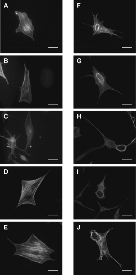
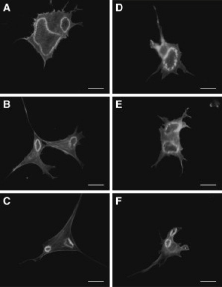
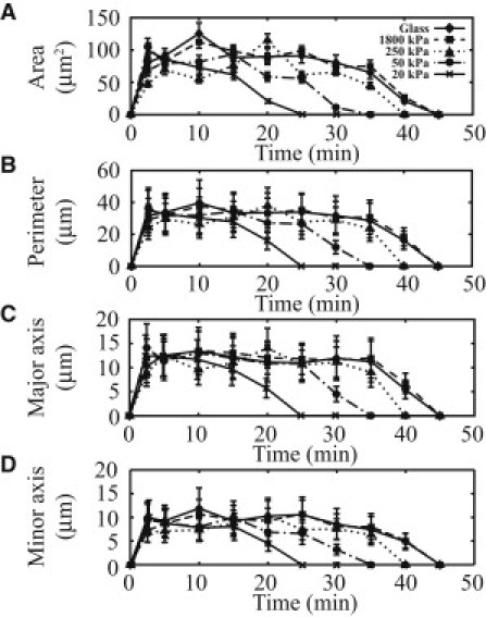
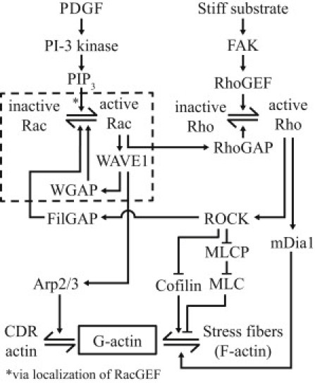
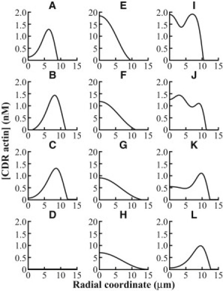
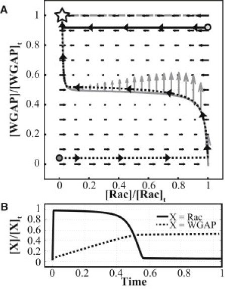
Circular dorsal ruffles (CDRs) are transient actin-rich ringlike structures that form on the dorsal surface of growth-factor stimulated cells. However, the dynamics and mechanism of formation of CDRs are still unknown. It has been observed that CDR formation leads to stress fibers disappearing near the CDRs. Because stress fiber formation can be modified by substrate stiffness, we examined the effect of substrate stiffness on CDR formation by seeding NIH 3T3 fibroblasts on glass and polydimethylsiloxane substrates of varying stiffnesses from 20 kPa to 1800 kPa. We found that increasing substrate stiffness increased the lifetime of the CDRs. We developed a mathematical model of the signaling pathways involved in CDR formation to provide insight into this lifetime and size dependence that is linked to substrate stiffness via Rac-Rho antagonism. From the model, increasing stiffness raised mDia1-nucleated stress fiber formation due to Rho activation. The increased stress fibers present increased replenishment of the G-actin pool, therefore prolonging Arp2/3-nucleated CDR formation due to Rac activation. Negative feedback by WAVE-related RacGAP on Rac explained how CDR actin propagates as an excitable wave, much like wave propagation in other excitable medium, e.g., nerve signal transmission.


Figures

Figure 1
Figure 1
Stiffness-based comparison of CDRs formed in cells. Cells stained for F-actin before (AE) and after (FJ) 5 min of PDGF stimulation. Cells were seeded on substrates with stiffnesses of (A and F) 20 kPa, (B and G) 50 kPa, (C and H) 250 kPa, (D and I) 1800 kPa, and on (E and J) glass. (Bars) 10 μm.
Figure 2
Figure 2
Time-based comparison of CDRs formed in cells seeded on (AC) glass and on (DF) 50 kPa PDMS substrates. The CDRs shown were taken of cells that had undergone (A and D) 10, (B and E) 20, and (C and F) 30 min of PDGF stimulation. (Bars) 10 μm.
Figure 3
Figure 3
Quantification of the size of CDRs observed in cells. The (A) area, (B) perimeter and (C) major and as well as (D) minor axes of the best fit ellipsoid to the CDRs are shown. (n = 3.) (Bars) One standard error.
Figure 4
Figure 4
Summary of events leading up to CDR formation from PDGF stimulation for generation of the complete model. The reduced model is constructed from the events (enclosed within the dashes). References for these events are provided in the text.
Figure 5
Figure 5
Simulations results for the effect of FAK concentration on CDRs. (A) Variation of radius of CDRs with time for different FAK concentrations. (B) CDR formation at T = 10 min and 20 min for [FAK] = 1 nM. Note that no CDR is observed at T = 40 min. (C) CDR formation at T = 10 min, 20 min, and 40 min for [FAK] = 10 nM.
Figure 6
Figure 6
Simulation results for the effect of WGAP and multiple PDGF receptor aggregates on CDRs. The variations of [CDR actin] in radial space in the presence of WGAP at (A) T = 10 min, (B) T = 20 min, (C) T = 30 min, and (D) T = 40 min after PDGF receptor activation at the origin are shown. The single peak in [CDR actin] is seen to travel away from the origin, illustrating a growing ring of CDR actin. In the absence of WGAP, similar plots are shown in panels EH. Note that without WGAP, the peak amount of [CDR actin] is elevated and centered at the origin, which translates to a patch of CDR actin that grows in size. Lastly, the variations of [CDR actin] after PDGF receptor activation at the origin and at radial coordinate of 7.5 μm is shown at (I) T = 2 min, (J) T = 4 min, (K) T = 8 min, and (L) T = 20 min. Note that only one peak is seen at later times, indicating that only one CDR actin ring is formed.
Figure 7
Figure 7
Phase diagram and time plots for Rac and WGAP, depicting the variation of Rac and WGAP with each other and in time, respectively. (A) Nullclines of F(x,y) = 0 (shaded line) and G(x,y) = 0 in (dashed line). (Arrows) Dynamics of Rac and WGAP (shaded arrows on the F(x,y) = 0 nullcline are scaled to 1000 times of the solid arrows). The stable steady state is indicated (star). (Solid curve) The rapid return of Rac and WGAP (from their values at the light-shaded dot) to their steady-state values when WGAP is only slightly decreased from its steady-state value. (Dashed curve) A typical course of excursion upon PDGF stimulation, where active Rac and active WGAP are both low, as indicated (dark-shaded dot). (B) Variation of Rac and WGAP with dimensionless time (with an initial state equivalent to that represented by the dark-shaded dot in panel A). WGAP attains the value of [WGAP]t at a much later time due to the slow dynamics of WGAP.

References

    1. Mellström K., Heldin C.H., Westermark B. Induction of circular membrane ruffling on human fibroblasts by platelet-derived growth factor. Exp. Cell Res. 1988;177:347–359. - PubMed
    1. Mellstroöm K., Höglund A.S., Lindberg U. The effect of platelet-derived growth factor on morphology and motility of human glial cells. J. Muscle Res. Cell Motil. 1983;4:589–609. - PubMed
    1. Tamura M., Iwamoto Y. The effect of platelet-derived growth factor on phagocytosis of cultured human trabecular cells. Exp. Eye Res. 1989;48:761–770. - PubMed
    1. Araki N., Egami Y., Hatae T. Phosphoinositide metabolism during membrane ruffling and macropinosome formation in EGF-stimulated A431 cells. Exp. Cell Res. 2007;313:1496–1507. - PubMed
    1. Arvidsson A.K., Heldin C.H., Claesson-Welsh L. Transduction of circular membrane ruffling by the platelet-derived growth factor β-receptor is dependent on its kinase insert. Cell Growth Diff. 1992;3:881–887. - PubMed

Publication types

MeSH terms

Substances