. 2011 Oct;3(10):1018-27.
doi: 10.18632/aging.100395.

Epigenetic-aging-signature to determine age in different tissues

Affiliations

Epigenetic-aging-signature to determine age in different tissues

Carmen M Koch et al. Aging (Albany NY). 2011 Oct.

Abstract

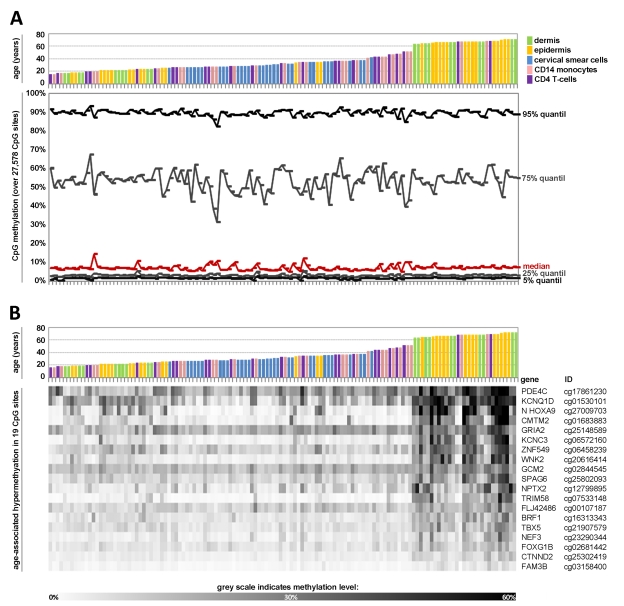
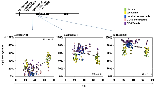
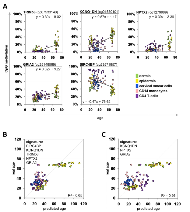
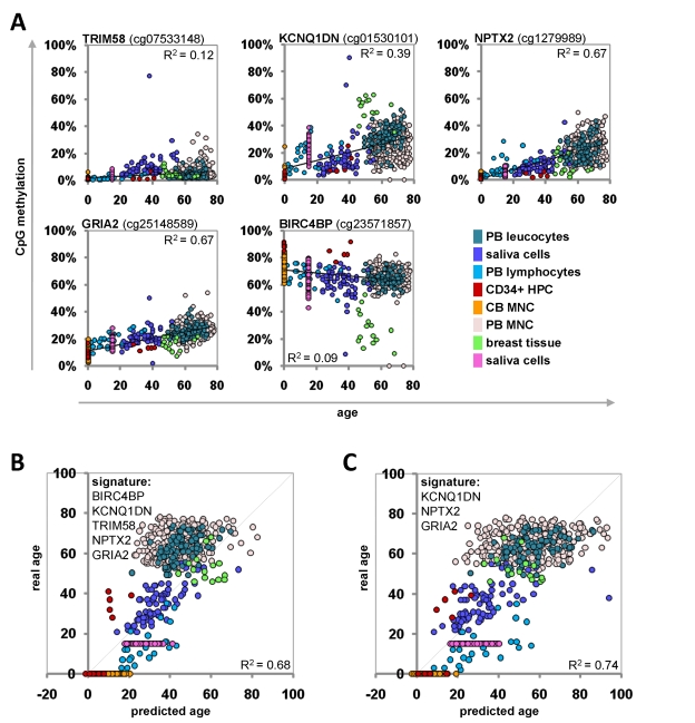
All tissues of the organism are affected by aging. This process is associated with epigenetic modifications such as methylation changes at specific cytosine residues in the DNA (CpG sites). Here, we have identified an Epigenetic-Aging-Signature which is applicable for many tissues to predict donor age. DNA-methylation profiles of various cell types were retrieved from public data depositories - all using the HumanMethylation27 BeadChip platform which represents 27,578 CpG sites. Five datasets from dermis, epidermis, cervical smear, T-cells and monocytes were used for Pavlidis Template Matching to identify 19 CpG sites that are continuously hypermethylated upon aging (R>0.6; p-value<10-13). Four of these CpG sites (associated with the genes NPTX2, TRIM58, GRIA2 and KCNQ1DN) and an additional hypomethylated CpG site (BIRC4BP) were implemented in a model to predict donor age. This Epigenetic-Aging-Signature was tested on a validation group of eight independent datasets corresponding to several cell types from different tissues. Overall, the five CpG sites revealed age-associated DNA-methylation changes in all tissues. The average absolute difference between predicted and real chronological age was about 11 years. This method can be used to predict donor age in various cell preparations - for example in forensic analysis.


Conflict of interest statement

The authors of this manuscript have no conflict of interest to declare.

Figures

Figure 1
Figure 1. Age-associated DNA-methylation changes at specific CpG sites across different tissues
Quantile analysis of beta-values in the 5 datasets of the training-group comprising: dermal cells (predominately fibroblasts), epidermal cells (keratinocytes), cervical smear cells (epithelial cells), and blood (monocytes and T-cells). The global distribution of DNA-methylation did not differ markedly with age or across different tissues (A). Pavlidis Template Matching (PTM) identified 19 CpG sites with age-associated hypermethylation (R > 0.6; p-value < 10−13) (B).
Figure 2
Figure 2. Age-associated hypermethylation and hypomethylation within KCNQ1DN
Schematic presentation of the promoter region with six CpG sites represented on the HumanMethylation27 BeadChip. Beta-values of three adjacent CpG sites were plotted against donor age.
Figure 3
Figure 3. Age-associated DNA-methylation changes at five CpG sites in the training-group
Methylation level of five selected CpG sites plotted against donor age. Regression coefficients and equations of linear regression are provided (A). Beta-values of the training group samples were used for the linear regression models to predict the donor age (R2= 0.65) (B). Alternatively, the signature was narrowed down to three CpG sites (R2= 0.56) (C).
Figure 4
Figure 4. Age-predictions with the Epigenetic-Aging-Signature for the validation group
Age predictions were tested with eight independent datasets. Beta-values of the five CpG sites were retrieved and plotted against donor age (A). The beta-values were used for the linear regression models of the training-set to predict the age of the donors based on 5 CpG sites (R2=0.68) (B) or 3 CpG sites (R2=0.74) (C) of the Epigenetic-Aging-Signature. PB = peripheral blood; CB = cord blood; HPC = hematopoietic progenitor cells; MNC = mononuclear cells.

References

    1. Ho AD, Wagner W, Mahlknecht U. Stem cells and ageing. The potential of stem cells to overcome age-related deteriorations of the body in regenerative medicine. EMBO Rep. 2005;6:35–38. - PMC - PubMed
    1. Schellenberg A, Lin Q, Schueler H, Koch CM, Joussen S, Denecke B, Walenda G, Pallua N, Suschek CV, Zenke M, Wagner W. Replicative senescence of mesenchymal stem cells causes DNA-methylation changes which correlate with repressive histone marks. Aging (Albany NY) 2011;3:873–888. - PMC - PubMed
    1. Fraga MF, Esteller M. Epigenetics and aging: the targets and the marks. Trends Genet. 2007;23:413–418. - PubMed
    1. Koch C, Suschek CV, Lin Q, Bork S, Goergens M, Joussen S, Pallua N, Ho AD, Zenke M, Wagner W. Specific Age-associated DNA Methylation Changes in Human Dermal Fibroblasts. PLoS ONE. 2011;6:e16679. - PMC - PubMed
    1. Gonzalo S. Epigenetic alterations in aging. J Appl Physiol. 2010;109:586–597. - PMC - PubMed

Publication types