

Prox1 transcription factor as a marker for vascular tumors-evaluation of 314 vascular endothelial and 1086 nonvascular tumors
- PMID: 22067331
- PMCID: PMC3288441
- DOI: 10.1097/PAS.0b013e318236c312
Prox1 transcription factor as a marker for vascular tumors-evaluation of 314 vascular endothelial and 1086 nonvascular tumors
Abstract
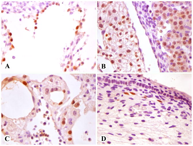
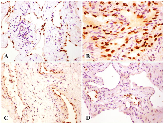
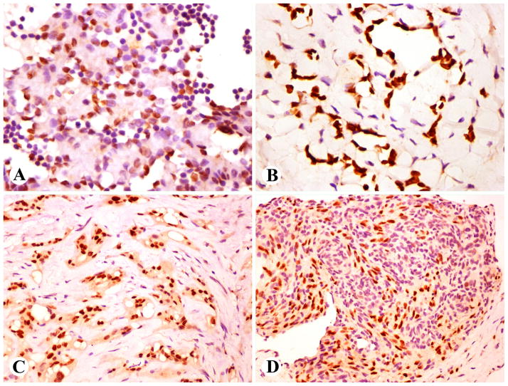
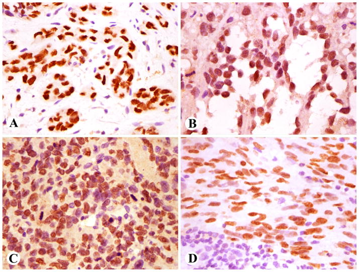
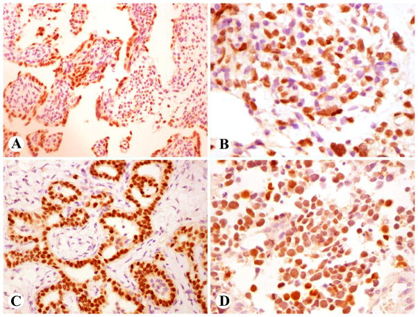
Prox1, a transcription factor important in the regulation and maintenance of the lymphatic endothelial phenotype, is consistently expressed in lymphangiomas and Kaposi sarcoma and has also been reported in Kaposiform hemangioendothelioma. However, information on its distribution in vascular tumors, such as angiosarcoma, is limited. In this study, we examined selected normal tissues and 314 vascular endothelial and 1086 nonvascular tumors to get an insight into the biology of these tumors and on potential diagnostic use of Prox1 as an immunohistochemical marker. In adult tissues, Prox1 was essentially restricted to lymphatic endothelia, with expression in subsets of pancreatic and gastrointestinal epithelia. However, it was also detected in embryonic liver and heart. Prox1 was consistently expressed in lymphangiomas, venous hemangiomas, Kaposi sarcoma, in endothelia of spindle cell hemangioma, Kaposiform hemangioendothelioma, and retiform hemangioendothelioma, and in half of epithelioid hemangioendotheliomas. It was present in most cutaneous angiosarcomas from different sites but was less commonly expressed in deep soft tissue and visceral angiosarcomas. In contrast, Prox1 was generally absent in capillary and cavernous hemangiomas. In positive hemangiomas and angiosarcomas it was coexpressed with podoplanin, another marker of the lymphatic endothelial phenotype. There was an inverse correlation with CD34 expression. The expression in mesenchymal nonendothelial neoplasm was limited. Prox1 was detected in 5 of 27 synovial sarcomas, specifically in the epithelia of biphasic tumors. Four of 16 Ewing sarcomas and 5 of 15 paragangliomas were also positive. All melanomas and undifferentiated sarcomas were negative. Among epithelial neoplasms, Prox1 was detected in 18 of 38 colonic carcinomas and 10 of 15 cholangiocarcinomas and in a minority of pulmonary, prostatic, and endometrial adenocarcinomas. The common Prox1 expression in angiosarcoma and its rare presence in nonvascular mesenchymal tumors make this marker suitable for the diagnosis of angiosarcoma and Kaposi sarcoma. However, the presence of Prox1 in some malignant epithelial tumors necessitates caution in applying Prox1 as a marker for vascular tumors. Common Prox1 expression in angiosarcoma may reflect the lymphatic endothelial phenotype in these tumors. Its patterns of expression in hemangiomas and angiosarcoma may be diagnostically useful and offer a new parameter in the biological classification of vascular tumors.
Figures
References
-
- Arai E, Kuramochi A, Tsuchida T, et al. Usefulness of D2-40 immunohistochemistry for differentiation between kaposiform hemangioendothelioma and tufted angioma. J Cutan Pathol. 2006;33:492–497. - PubMed
-
- Burke Z, Oliver G. Prox1 is an early specific marker for the developing liver and pancreas in the mammalian foregut endoderm. Mech Dev. 2002;118:147–155. - PubMed
-
- Dadras SS, Skrzypek A, Nguyen L, et al. Prox-1promotes invasion of kaposiform hemangioendotheliomas. J Invest Dermatol. 2008;128:2798–2806. - PubMed
-
- Dudas J, Papoutsi M, Hecht M, et al. The homeobox transcription factor Prox1 is highly conserved in embryonic hepatoblasts and in adult and transformed hepatocytes, but is absent from bile duct epithelium. Anat Embryol (Berl) 2004;208:359–366. - PubMed
Publication types
MeSH terms
Substances
Grants and funding
LinkOut - more resources
Full Text Sources
